Anatomy of the Lower Back

hip_musculature_spinal_support

(Part 1 of 2: Muscular Skeletal System)

If you have practiced yoga lately, chances are that you sat on the floor for a little while. This is a very healthy activity that every human should probably practice regularly for the strength of the pelvic floor muscles, and to allow the inner thighs and hips to relax. You can always work your way into it with blocks, props, cushions, pillows; you can do it while watching TV. It is good for releasing the muscles in the lower spine which have a strong connection with the hips, pelvic floor, abdominal muscles, lower back, and lower organs, including the sex organs and excretion organs; it’s good for all that important stuff.

In this article, I will speak specifically about the lower back and the anatomical features that you will want to be aware of as you practice yoga. If you have any of the following symptoms, you may want to begin a restorative yoga practice to assist in the alleviation of your pain, as well as begin to sit on the floor regularly. If it is too painful to start, contact a specialist or something like that, here are some symptoms of dysfunction:

  • Pain and stiffness in the back.
  • Pain in the buttocks and the legs, often in the back of the thigh.
  • Pain that worsens when bending, stretching, coughing, or sneezing.
  • Sciatic Nerve pain (pain in the hips, or back of your legs that shoots down the leg)

The lower back is really composed of three areas of the body: the lower spine, the hips and tailbone, and the abdomen. Since the spine is encircled by musculature, the abdomen, spinal muscles, and hips are all integral aspect of maintaining a healthy lower spine and therefore lower back.

Here is a depiction of the skeletal frame with the lower back in red: lumbar_region from wikipedia

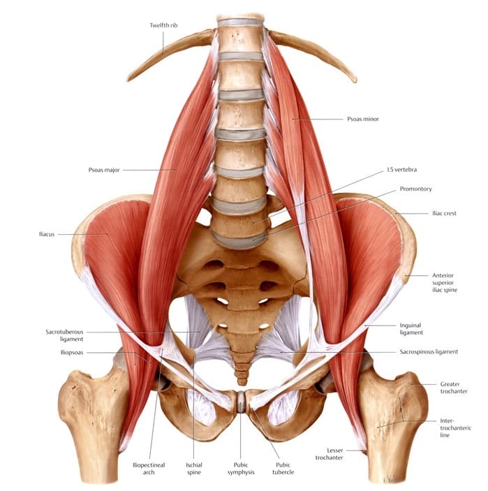
You can see the there is a lot of big bone support at the base of the spine you will know from your own body that your hips provide the support for the lower spine. The ribs and upper legs have a tremendous amount of connection with the lumbar region of the spine and are the primary support structures in providing space for the lower back and lowest organs. Here is a very detailed depictions of the inner hips muscles and lower spine: hip_musculature_spinal_support

This is a depiction of the primarily of the psoas muscles and illiacus muscles. The psoas is a primary muscle group that moves the trunk Gray's Quadratus Lombrumand is greatly affected by sitting habits. It lines the font of the spine and inserts separately into each vertebral process up to the T12 in most people. The illacus muscles line the insides of the hips and connect with the psoas at the insertion point of the lesser trochanter of the femur. Both the psoas and the QL run along the lumbar spine to the trunk, the QL going posterior to the spine and the psoas anterior to the spine, bone are connected to the transverse spinal processes. The muscles work together to move the trunk, along with the muscles of the abdomen. The psoas and the QL muscles are the primary muscles of the lower back, so we’ll come back to them.

There are a few more groups of muscles to pay attention to, but other primary muscle group to consider when talking about the lower back is the abdominals. Your abdominals provide frontal support for the spine, but in addition to the abdomen and primary lower back muscles of the Psoas and Quadratus Lumborum, the diaphragm, obliques, serratus muscles, pyramidalis muscles, levatores costarum, subcostal muscles, transverse thoracis muscles, and intercostal muscles play roles in the alignment of the lower spine. The final, possibly most under looked muscle is the latimus dorsi, which runs all the way along the back of the spine up to the shoulders. We can go over most of them as accessory breathing muscles, which is an action largely affected by the lower spine. It sounds like a lot of muscles because there are lots of muscles that are connected to your lower back. Let’s break it into pieces to see how it works.

Lets start at the top and work out way down the body, so lets start with the shoulders. The serratus muscles, obliques, levatores costarum, costal muscles and subcostal muscles all play a role in spinal alignment at the shoulder level. The subcostal muscles are the subcostal_muscles_ depictioninnermost, being inside of the rib cage, and surrounds the diaphragm along the ribs. The intercostal muscles are just superior, or further outside than the subcostal muscles. The levatores costarum run along the back of the spine on the outside of the rib cage, "Levatores costarum" by Uwe Gille - modified from Image:Gray389.png. Licensed under Public Domain via Wikimedia Commons - http://commons.wikimedia.org/wiki/File:Levatores_costarum.png#/media/File:Levatores_costarum.pngproviding even more support for the spine, which you can see act in opposition to the interlacing rib muscles. Notice the spinal erectors and spinalis muscle groups. You can also see how the muscles interweave with the spine and ribs, making breathing a full body movement. This is why forward folds are so effective at releasing the muscles the support the spine, so that they can stretch and relieve tension. This part of the reason why breathing in yoga can help to align the spine, and why spinal alignment and breathing have a close relationship. The obliques are a portion of the abdomen that you can read more about here. The serratus anterior is the another muscle to consider, which is also known as the punching muscle, as it pulls the shoulder blades forward. This is an extremely strong and useful muscle in yoga that supports you in handstands, forearm stands, and headstands in specific variations with proper alignments. and keeps the shoulders aligned, which then keeps the spine aligned. Like links on a chain. The final muscles to look at is theLatissimus_dorsi latimus dorsi, a muscle that runs from the lower back to the outside of the shoulder blades. The muscle connects the arms to the lower back, so can be really important for golfing, blowing, javelin throwing, or boxing. Anything where you are using your hips to power the upper body. These muscles can be easily overlooked in sun salutations, especially downward dog, which can allow the spine to hyperextend. This muscle is also more active in the elbow close push up, allowing the serratus anterior and lats to move the spine up from chaturanga into downward dog. Hollowing your armpits in plank/chaturanga/down-dog will likely activate and strengthen your lats, but its good to have a second pair of eyes on your alignment, so if you have questions find a local studio or teacher and ask them about your down dog. I’m sure they will be thrilled to answer your questions.

The last muscles to consider are the diaphragm, the obliques, the

Diaphragm
Diaphragm

pyramidalis muscles, and the transverse abdominus. The final piece of the puzzle is the rectus abdominus, which acts in direct opposition to the Psoas and QL muscles. The pyramidalis muscles are tiny triangles below the lowest layer of the rectus abdominus, and they form muscles just above the genitals. The diaphragm forms the inner musculature needed to move the ribs as the lungs expand.

abdomen image from http://www.usra.ca/The obliques line the outsides of the torso at the bottom of the rib cage, and all four layers of abdominals meet at the linea alba and run down to the pyramidalis muscles. You can see the lowest layer of the abdominals, the transverse abdominus, which acts as a kind of weight belt to support the lower spine when heavy lifting with the back, or squatting. It works with the psoas and QL to keep the trunk stable. The rectus abdominus acts in opposition to the QL and psoas, which forms a kind of push-pull system for you to lean forward and back, to squat, and to jump. Think of them as working against each other, but really they work in unison to support your spine. A great way to feel all of these muscles is to do burpees and/or sun salutations.

That wraps up the muscular and skeletal portions of the anatomy of the lower back. Please check back in about a week for the second section where I discuss nerves, organs, blood vessels, and fluid distribution, and if you are looking for something a bit more entertaining, you can check out the WANDERER series, I am working on part 13 right now and should have it out in a few days. Thanks for reading, would love to hear any questions or feedback


Lumbar Spine Anatomy (part 3: Lumbar|Lower Spine)

lumbar spine_vertebrae

Lumbar Spine

The lumbar spine is the lowest five of the twenty-four vertebrae of the spine, which descend vertically from the bottom of the ribs to the top of the hips and pelvis. Structurally, the lumbar spine is the foundation for the body’s movement and supports the weight of the upper body as it moves through space. It is prone to injury because it is where the most weight-bearing and movement of the body takes place.

The lowest and widest bone of the column is the L5, with large amount of ligamentation towards the fused sacrum and the posterior, superior portion of the iliac crest, or hip plate. As one descends down the vertebrae, the pedicles of each bone get longer and deeper as the spinal cord widens. The vertebrae have 5 wide fins, called processes, two transverse processes, two articular processes, and the spinous process at the very rear of the vertebrae. The spinal cord passes through the vertebral foramen which is the chamber formed by the processes at the back of the bone.

The Ligaments of the Lower Back

There are multiple ligaments connecting each vertebrae and interlacing down into the sacrum. The lumbar portion of the spine is the least able to twist as it is primarily a support structure for the body. Each bone of the Lumbar is wider and deeper than the last, allowing for less movement and more stability as you descend down to the pelvis. The backsides of the vertebrae interlace with ligaments and tendons that stabilize the trunk. The psoas muscles run up the lumbar vertebrae on each side up to the L1. The Lumbar is the third and final curve in the spine, with ligaments interlacing down into the sacrum and up into the thoracic spine and ribs.

The Nerves of the Lumbar Spine

The lumbar spine is particularly important for the nervous system; it is the starting point for the nerves of the legs and lower body and contains a plexus of nerve connections that extend up the spinal spine_nervescord, and down into the legs and lower organs. This is a pivot place for alignment of the entire body; oftentimes nerve pain originates from mis-alignment in the lower back. The fins and processes at the back of the bones protect the nerves, but can be somewhat easily mis-aligned to pinch nerves in the legs and hips. This is why hip opening should be done slowly and why it is important to keep the sitting muscles (QL, spinal erectors, obliques) engaged and lengthening throughout hip opening.

Muscles of the Lumbar Spine

The muscles that interlace in the Lumbar spine are intricate and not usually appreciated by the term spinal erectors. The spine seems to be perfectly capable of strengthening itself to stay erect, if you engage the obliques, transverse abdominus, the muscles that run up the spinal vertebrae, and the serratus posterior inferior muscles connecting to the lumbar vertebrae from the ribcage.

intrinsic-back-muscles

That concludes the final piece of anatomy of the spine. Hope you enjoyed the series, parts 1 and 2 are available by clicking on the numbers. Let me know if you want me to write about anything else in the spine!


Anatomy of the Spine (part 2: Thoracic)

The twelve vertebrae in the middle of your back comprise the thoracic spine, below the 7 cervical and above the 5 lumbar vertebrae. Each vertebrae connects ribs in the front of the body to form the rib cage which connects to the spine between the thoracic vertebrae. The thoracic spine could be considered the most versatile, though the cervical is capable of greater rotation and the lumbar provides more support for the skeletal structure. It also protects and provides nerve supplies for the essential organs including the heart, liver, kidneys, stomach, lungs, and intestines inside of the rib cage. This area of the spine is one of the most important and is also one of the most unusual skeletal structures seen in nature (due to being the middle of the unique, three curve spine).

The thoracic discs are particular, allow for certain movements spineand more flexibility and rotation at the superior portion.You can see the differences between the cervical discs and lumbar discs on the right. The curvature of the spine is very apparent and is what allows for human biped movement (apes use their hands on the ground because of the singular curve in their spine; this hints towards the progression of the human spine. You can also see the significant rotation that the thoracic vertebrae allow for, giving the skeletal structure mobility above the lumbar spine.

The Thoracic spine is one of the most important nervous gateways in the body, creating the gateway between the peripheral nerves spinal nerve mapof the upper body and the central nervous system. The peripheral nerves begin to leave the spinal nerves at the ganglion nerves, or connective nerve clusters, for the arms where the thoracic spine begins; the nerves then cascade down to connect all of the ribs, until the lumbar spine connects to the lower body and lowest abdominal organs. Essentially, the nervous system controls the entire upper body through the thoracic nerve connections and the biggest vital organs sit right in the middle of these nerves. nervesThe thoracic spine connects directly to the rib cage, making it one of the most important protective structures of the human

autonomic nervous systemskeleton. The heart has nerves that extend up into the neck (think about the thyroid’s control over the heart) and is largely connected to the middle vertebrae of the spine. The lungs also share these thoracic nerves and nestle the heart; they are meant to provide the heart with support and surround it. The stomach, liver, pancreas, small intestine, and adrenals share the lower portions of the thoracic nerves, while the large intestine has connections to both the lumbar spine and the thoracic spine. thoracic muscles

There are many different muscles of the thoracic spine and middle back because the thoracic spine is the gateway to control center of the upper body, which is where the majority of movement takes place (the hands and fingers are finely tuned and the shoulder is a durable and versatile joint). The vertical muscles that extend up the spine and interconnect vertebrae are the deepest layer of muscle and the diaphragm (the breathing muscle) is also included in this musculature.

deep thoracic musclesThe deepest muscles of the spine interconnect the vertebrae to ensure structural stability. There are also fascia that extend up and down the length of the spine, known as the vertical erectors (they help humans to stand up straight). vertical spinal erectors You can see them detailed on the left. There is also significantly denser ligamentation towards the inferior portion of the spine, leaving the upper portion more mobile and connected with muscular tissue. The spinal cord itself is completely protected and supported by these muscles, allowing for the extensive movements that humans are capable of.

This concludes the second part of three about the human spine and its anatomical functionality. If you have any questions, feel free to comment, but stay tuned for part 3 where we will dive into the lumbar portion of the human spine.

Anatomy of the Spine (Part1: Cervical)

Cervical Spine

The human spine has 24 vertebrae and is usually separated into 3 sections: the Lumbar curve, the Thoracic curve, the Cervical curve. I will be doing a three-part series for the spine, as the spine as a whole is too complicated to summarize effectively in one post. We will move from top to bottom and each post will get longer as we move closer to the base of the spine. For now, we will start with the seven Cervical vertebrae which allow for the greatest rotation and support the neck, skull, and brain. These are the most mobile vertebrae and have extraordinarily complex neural, venous, and muscular connections and passageways.

Cervical Vertebrae 1

C1 (C1-C7 are cervical, C1 being on top) is the highest spinal vertebrae and is also known as the Atlas. This is drawn from Greek mythology as a metaphor because the atlas supports the globe of the head. This vertebrae is special as you can see from its shape; the brain stem extends into the vertebrae at the same time as the vertebrae has lots of room to tilt forward, back, and to the sides. The atlas is also fused with the Axis (C2) and has no body as a

Posterior ligaments of C2

result. The Axis is the seat upon which the Atlas rotates and provides support to circularly rotate the head. These first two vertebrae are particularly significant because the brain stem connects the spinal cord at the base of C2 to the brain.

C3 through C6 are somewhat uniform and share many characteristics. In order to move forward, we need some vocab work and general characteristics of vertebrae to be able to compare them.

Vertebral Characteristics : A typical vertebra consists of two Vertebral arch labelledessential parts— ananterior segment, the body, and a posterior part, the vertebral or neural arch; these enclose a foramen, the vertebral foramen. The vertebral arch consists of a pair of pedicles and a pair of laminæ, and supports seven processes—four articular, two transverse, and one spinous.

body – The thick connecting structure supporting the bone
vertebral arch – The extensions from the body which connect to each other through ligaments and muscle tissue
pedicles – The two short process which connect the arch to the body of the vertebrae
laminae – two broad plates extending from the pedicles
process – anatomical terms for an extension, or outgrowth of tissue from a larger body
transverse processes – project up and down from where the lamina meets the pedicles. In the cervical spine this is pierced by the transverse foramen, which is discussed below
spinous processes – the fin like posterior tip of the vertebrae that extends back from the lamina
articular processes – these connect the vertebrae at the junction of the lamina and pedicles, form the links of the spine
tubercle – describes a round nodule, small eminence, or warty outgrowth found on bones or skin

Now that we have a decent idea about the characteristics of vertebrae we can look in detail at the cervical vertebrae. The first six vertebrae are wider than they are long and they overlap with the front of the vertebrae below. The Laminae are narrow, but widen on the inferior side, so each vertebrae is wider at the bottom than top make the lower vertebrae wider than the upper. The spinous processes are short and bifid (split), with one end being longer than the other. Articular pillars are formed by the superior and inferior articular processes that have fused and link the vertebrae together. The transverse processes are pierced by the transverse foramen, which gives passage to the vertebral arteryvertebral vein, and a plexus of sympathetic nerves. The seventh foramen lacks the artery, but contains the vein and sympathetic nerves.

The seventh cervical vertebrae is fairly unique. It is called the Vertebra prominens and has a long and palpable spinous process Cervical Nervesthat isn’t split and that you can see in about 70% of people, hence its name. It’s considerably bigger spinous process gives it more support for veins and nerves which start to bundle heavily and for the muscles that extend horizontally in the shoulders and down the arms. See more detail on the nervous connections to the right.

The veins and arteries of the cervical vertebrae run parallel to the left and right of the spinal column. It is a complex wiring that interlace with each vertebrae.

Cervical Veins

The muscles of the neck are detailed in a prior post here. Stay tuned for the Thoracic spine, the middle 12 vertebral columns and how they function within the body.