anatomy

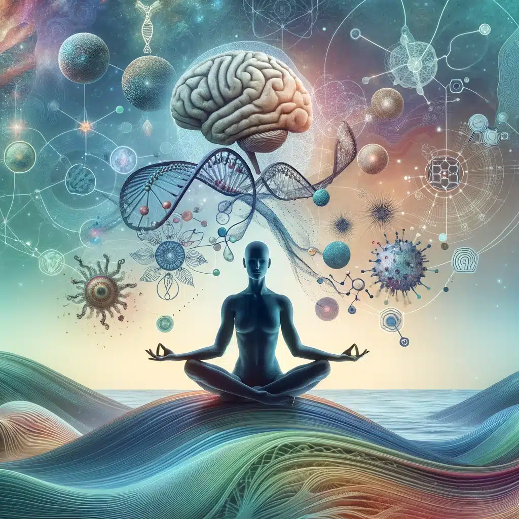
The Science of Serenity: Yoga’s Impact on the Nervous System and Hormonal Balance

Introduction

Over the past 10 years, I have been attempting to understand and adapt the ancient practice of yoga. I travelled to Mysore to study ancient yogic traditions through the lens of modern science. It has also helped me to understand what brought me to yoga originally. Over the course of teaching thousands of students yoga, I can confirm that the benefits of yoga are tremendous and very much understated in modern society. It’s simple; health is declining because it isnt valued. The practice of yoga allows for an individual to realize fascinating and comprehensive benefits for human health and to redistribute their system of valued. Yoga is a way of philosophy. This article will delve into the anatomical and physiological underpinnings of yoga, specifically its profound effects on the nervous system and hormonal balance, to illuminate how these practices foster positive mental health.

Yoga and the Nervous System

The Parasympathetic Nervous System and Yoga

Yoga’s ability to activate the parasympathetic nervous system (PNS), often referred to as the “rest and digest” system, is a cornerstone of its mental health benefits. According to a study published in the Journal of Alternative and Complementary Medicine, practices such as gentle yoga poses, meditation, and pranayama (breath control) significantly increase vagal tone, an indicator of parasympathetic activity, leading to relaxation and stress reduction (Streeter et al., 2010).

Just sitting still and breathing deeply can lower your cortisol levels significantly. I can’t tell you how many people miss out on this in favor of the “go” mentality. To relax, you have to stop; sometimes.

Neuroplasticity and Mindfulness Practices

Research in Frontiers in Human Neuroscience outlines how consistent yoga practice contributes to neuroplasticity, the brain’s ability to form new neural connections throughout life. This adaptability is enhanced by yoga’s mindfulness component, promoting cognitive flexibility and resilience to stress (Gard et al., 2014).

These findings collectively suggest that yoga can play a pivotal role in mental health interventions. By enhancing neuroplasticity and fostering a mindful approach to mental health, yoga offers a holistic and effective strategy for managing anxiety and depression, supporting its integration into mental health treatment and wellness programs for addicts and/or abusers.

Meditation and Brain Structure: Research has shown that meditation, a key component of yoga, can lead to changes in the brain’s structure, particularly in areas associated with memory, sense of self, empathy, and stress. For instance, a study that found increased gray matter density in the hippocampus, known for its role in learning and memory, in individuals who engage in mindfulness meditation.

Breath control and pranayama practices influence the autonomic nervous system, shifting the balance towards the vagus nerve, which helps promote relaxation and reduce stress levels. This shift is associated with reduced cortisol levels, a marker of stress, thereby potentially reducing anxiety and depressive symptoms.

lakeyoga_Elliot&Mandy

Yoga for Hormonal Balance

Cortisol and Stress Response

A pivotal study in Psychoneuroendocrinology demonstrated that regular yoga practitioners exhibit lower levels of cortisol, the stress hormone, compared to non-practitioners. This suggests that yoga can modulate the body’s stress response system, leading to improved mental health outcomes (West, Otte, Geher, Johnson, & Mohr, 2004).

Yoga’s Effect on the Endocrine System

Yoga’s impact extends to the broader endocrine system, which regulates hormones. The Indian Journal of Physiology and Pharmacology reported that specific yoga poses and sequences can stimulate or soothe various glands, promoting hormonal balance. This equilibrium is essential for mood regulation, stress management, and overall mental well-being [1]. Studies have indicated that mindfulness practices can reduce rumination, a significant factor in the development and maintenance of depression. By fostering a non-judgmental awareness of the present moment, yoga helps individuals break the cycle of negative thought patterns.

Practical Applications

The practice of asana can optimize the nervous system and hormonal functions. For example, inversions like downward dog or headstand can rejuvenate the endocrine system, while slow, mindful movements in poses such as child’s pose or seated forward bend activate the vagus nerve, fostering a state of calm.

Yoga for Substance Abuse Disorders

Yoga has been increasingly explored as a complementary intervention strategy in the treatment of substance use disorders (SUDs), with several studies highlighting its potential benefits. Research demonstrates that yoga practices, including Hatha yoga, Sudarshan Kriya yoga, and various breathing and meditation exercises, can have positive effects on individuals struggling with substance use, including tobacco, alcohol, or opioids.

Incorporating Scholarly Insights into Daily Practice

Understanding the science behind yoga empowers practitioners to tailor their practice to specific mental health goals. When you are stressed, you can meditate. By integrating these scientifically backed techniques into regular practice, you can control your own health. There is a lot of research left to be done; but there is some evidence to support yoga as an effective therapy for anxiety and depression. Obviously these are hard things to measure; not only are depressions and anxiety subjective, but the way each individual experience yoga is different because of different use histories in the body from sports, etc.

Conclusion

These findings collectively suggest that yoga, through its multifaceted practices of breathing, contorting, and physical rigor can ameliorate mental health. The confluence of yoga with modern scientific research offers compelling evidence of its benefits. The key to further research will be underpinning its exact efficacy in enhancing mental health. So far, the breathing techniques have yielded some of the most widely acknowledged clinical data. As we continue to explore the depths of yoga’s therapeutic potential, it becomes clear that this ancient practice holds timeless relevance in our quest for psychological well-being in the contemporary world. During our lifetime, we might learn why!

References (read the first one!)

  1. Bhavanani, A. B., Madanmohan, & Sanjay, Z. (2012). Understanding the Science of Yoga. The Indian Journal of Physiology and Pharmacology.
  2. Gard, T., Noggle, J. J., Park, C. L., Vago, D. R., & Wilson, A. (2014). Potential self-regulatory mechanisms of yoga for psychological health. Frontiers in Human Neuroscience.
  3. Streeter, C. C., Gerbarg, P. L., Saper, R. B., Ciraulo, D. A., & Brown, R. P. (2010). Effects of yoga on the autonomic nervous system, gamma-aminobutyric-acid, and allostasis in epilepsy, depression, and post-traumatic stress disorder. Journal of Alternative and Complementary Medicine.
  4. West, J., Otte, C., Geher, K., Johnson, J., & Mohr, D. C. (2004). Effects of Hatha Yoga and African Dance on Perceived Stress, Affect, and Salivary Cortisol. Psychoneuroendocrinology.
  5. Namrata Walia, Jennifer Matas, Acara Turner, Sandra Gonzalez and Roger Zoorob -The Journal of the American Board of Family Medicine

The Science of Serenity: Yoga’s Impact on the Nervous System and Hormonal Balance Read More »

artiphoria-prana-entering-the-body

The Anatomy of Breath: A Yogi’s Guide

Artiphoria.ai AI generated image

To sustain life, a body must produce sufficient energy through aspiration. Breathing is perhaps the only system of the body that is both autonomic and conscious depending completely on the awareness and focus of the breather.

Breathing through the nose, all the time, is part of the true yogi’s path. I can remember 6 months into practicing yoga, I attained the ability to breathe through my nose and it complete changed my yoga practice and my life. I got hooked on the feeling of yoga (call it a healthy addiction) and never looked back.

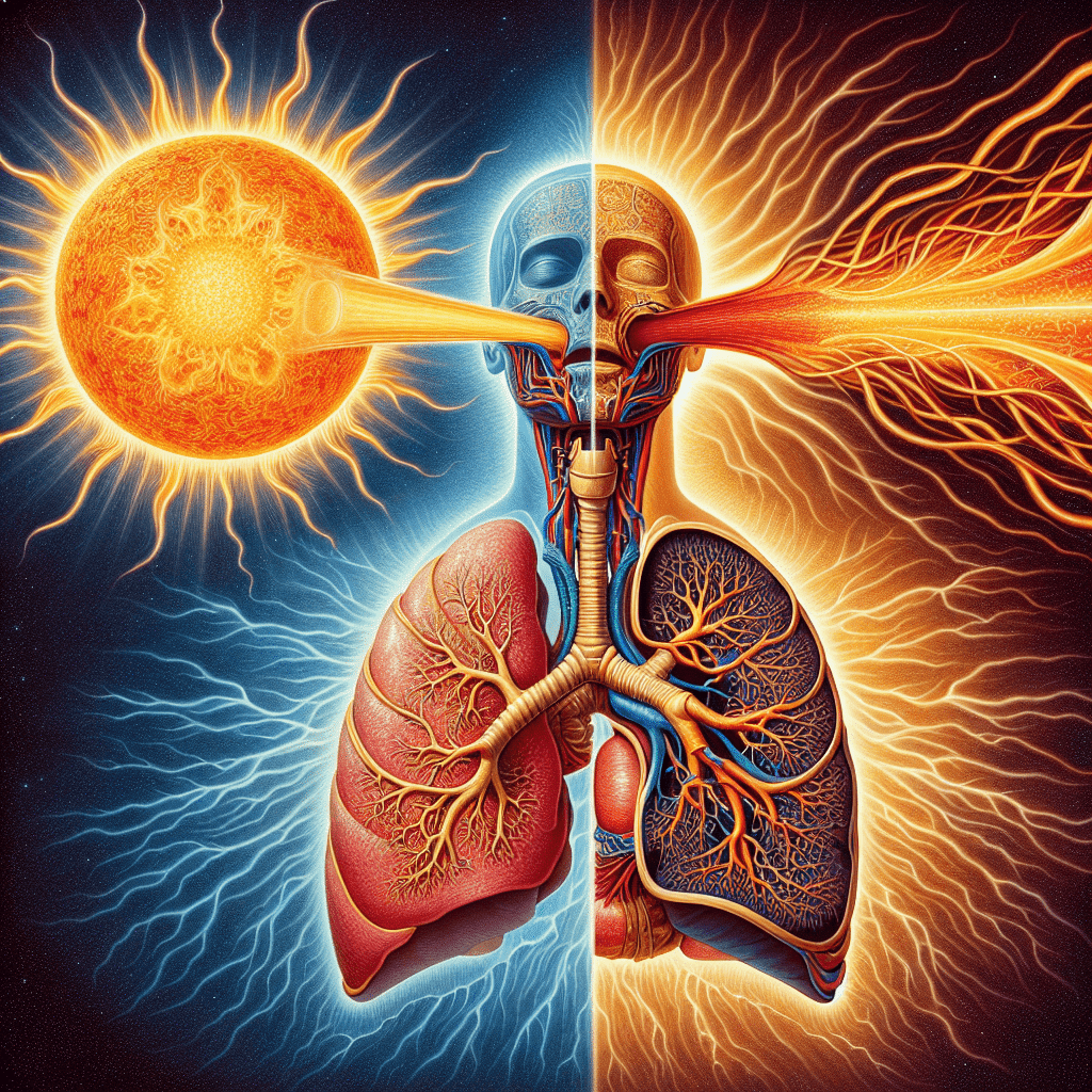
In Yoga, the energy of breath is called prana (प्राण, prāṇa) which can be described as solar wind in the atmosphere, or liquid light[6]. Through ventilated aspiration, the yogi ingests the prana into the nervous system. In Hindu literature, Prana is described as originating from the Sun and connecting the elements through the Chakras of the human nervous system and conscious awareness.

Yousun Koh

The nervous system is completely dependent on your breathing to function: The parasympathetic system slows your breathing rate. It causes your bronchial tubes to narrow and the pulmonary blood vessels to widen. The sympathetic system increases your breathing rate. It makes your bronchial tubes widen and the pulmonary blood vessels narrow.[4] This process of is also known as the “fight or flight” response. This happens through ventilation, or respiration as the body mobilizes itself to a threat. However, this system is over-active in our cultures because of our stress responses to non-life threatening stimuli. It is healthier for a human to regularly breathe through the nose.

The Nasal Cavity

“The function of the nasal cavity is to warm, moisturize, and filter air entering the body before it reaches the lungs.[1]” Here are the additional benefits:

Nose breathing is beneficial primarily because it allows your nasal cavities to:[2]

msdmanuals.com/home/lung-and-airway-disorders
  • reduce exposure to foreign substances.
  • humidify and warm inhaled air.
  • increase air flow to arteries, veins, and nerves.
  • increase oxygen uptake and circulation.
  • slow down heart rate.[3]
  • improve lung volume.
  • help your diaphragm work properly.

In essence, hairs and mucus lining the nasal cavity help to trap dust, mold, pollen and other environmental contaminants before they can reach the inner portions of the body and the lung’s organic tissue. Air exiting the body through the nose returns moisture and heat to the nasal cavity before being exhaled into the environment.[1] The mouth, also known as the oral cavity, is the secondary external opening for the respiratory tract. The mouth is mostly for filtering in eating and drinking.

Oblique muscles (accessory breathing muscles)

Focus on the Exhale

Nasal exhalations are an extremely important focus in yoga. The muscles in your chest and abdomen tighten or contract to create a slight vacuum around the lungs. This causes air to flow in. When you exhale, the muscles relax and the lungs deflate on their own, much like an elastic balloon will deflate if left open to the air. The lungs are extremely flexible sacks of tissue that have the ability to expand and contract.

References:
Yousun Koh
  1. InnerBody.com
  2. Dentallogictruro
  3. YogaU Online
  4. National Institute of Health (Govt)
  5. Himalaya Institute – science of breath
  6. NASA – Solar Wind
  7. kenhub.com
  8. Sivananda

The Anatomy of Breath: A Yogi’s Guide Read More »

Henry Gray – the Father of modern anatomy

By H. Pollock - [1] [2], CC BY 4.0, https://commons.wikimedia.org/w/index.php?curid=1458730

Henry Gray was an innovator and disruptor in the medical field with his surgically precise incisions and methodically meticulous explorations of the human anatomy. I love his work and have used the depictions from his book for much of my anatomy articles due to their lack of copyright.

Gray was born in Belgravia, London in 1827 and spent most of his life in London.

His book on human anatomy, Gray’s Anatomy, is still regarded as the anatomical bible of the scientific world. He was extremely precise and through his experience making painstakingly exact incisions and methodically mapping the physical anatomy of the human body. While still a student, Gray received the triennial prize of Royal College of Surgeons for his essay The Origin, Connexions and Distribution of nerves to the human eye and its appendages, illustrated by comparative dissections of the eye in other vertebrate animals.

The following year, at the age of 25, Gray won another prize for his essay on the Spleen(this is a fascinating read when you have time), and was elected a fellow of the Royal Society.

In 1858, Gray Published his first anatomy book of 750 pages and hundreds of figures by his friend Henry Vandyke Carter. They met at St. George’s School of Medicine. At first, when Gray wrote his essay on the spleen, Carter thought Gray was a snob. Over time and as they worked together more their respect for each other grew, largely due to their seriousness and commitment to the field of medicine. Initial conflict occurred with respect to the payment that Carter received from Gray for his designs, being in need of money, and that he judged inadequate for his commitment.[5]

The images from these two scientific heroes are timeless. And luckily for me, they are copyright free due to their age and remain some of the most powerful anatomical depictions. I use many of them in my anatomical articles on this website.

Henry Gray’s Death

Gray was struck by attack of confluent smallpox, a most deadly kind of disease. On 13 June 1861, the day he was to appear for an interview as a final candidate for a prestigious post at the St. George’s Hospital, he died at the age of 34.[3] He was buried at Highgate Cemetery.[4][5] Gray had been vaccinated against smallpox as a child with one of the early forms of the vaccine.[6]

He is assumed to have been infected due to his passionate care giving for his ten-year-old nephew, Charles Gray, who did recovered from the deadly disease.

The Style of the Book

had a major influence on science that has since but irreplaceable. It is reminiscent of the evolution of art from Classicism to Hellenism in Ancient Greek art, throughout the Renaissance and afterwards, in that the accurate and precise portrayal of the human form was the primary goal. Read more about the stylistic evolution of anatomy here.

You can buy a cool version of Gray’s Anatomy Here.
References:
  1. Henry Gray’s Anatomy of the Human Body
  2. Henry Gray on the Structure and Use of the Spleen
  3. Henry Gray Wikipedia Page
  4. Vandyke Carter (Henry) Wikipedia Page
  5. Royal Society Wikipedia PageRoyal Society Website
  6. The Making of Gray’s Anatomy
  7. Internet Archive of Gray’s Anatomy
  8. Style and Non-style in Anatomical Illustration

Henry Gray – the Father of modern anatomy Read More »

Femur

Femur Bone Anatomy: Pillars of Support for the Human Skeleton

Introducing the most Massive and Strong (in most ways) Bone in the Human Body

There are 62 bones in the legs: 10 trunk/hip bones, 14 ankle bones, and 38 foot bones. The femur (thigh) is the largest and strongest of these bones. Most land mammals capable of jumping also have femur bones, also lizards, frogs, and other tetrapod vertebrates. Its length on average is 26.74% of a person’s height, a ratio found in both men and women and most ethnicities with only restricted variation.

"80 - Pelvic bones with sacrum and femur" by Knowledge Collector is marked with Public Domain Mark 1.0. To view the terms, visit https://creativecommons.org/publicdomain/mark/1.0//?ref=openverse.

5 Interesting Femur Bone Statistics

  1. The femur is, on average, about 1/4 of a person’s height. It’s remarkably strong and can withstand forces of up to 1,800 to 2,500 pounds (800 to 1,100 kilograms) of pressure, making it one of the strongest bones in the body.
  2. The Femoral neck sits at a 125 degree angle
  3. Vehicular accidents are the primary cause of breakage
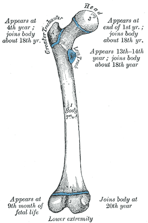
  4. During growth in childhood and adolescence, the proximal end of the femur (the femoral head) has a growth plate, known as the epiphyseal plate. This growth plate allows for longitudinal growth and helps determine a person’s final height when it closes.
  5. Forensic anthropologists often use the femur bone to estimate the age of an individual based on the degree of fusion of the epiphyseal plates, which can help in identifying human remains. It is heavily used in archaeology.

The Greater Trochantergreater_trochanter_grays

The Great Trochanter is a large, irregular, quadrilateral eminence on the upper portion of the femur bone. This portion of the bone has several, extremely important muscle insertions for the thigh and hip bones:

The lateral surface, quadrilateral in form, is broad, rough, convex, and marked by a diagonal impression, which extends from the postero-superior to the antero-inferior angle, and serves for the insertion of the tendon of the gluteus medius.

human_ape_femurs

Above the impression is a triangular surface, sometimes rough for part of the tendon of the same muscle, sometimes smooth for the interposition of a bursa between the tendon and the bone. Below and behind the diagonal impression is a smooth triangular surface, over which the tendon of the gluteus maximus lies, a bursa being interposed.

The medial surface, of much less extent than the lateral, presents at its base a deep depression, the trochanteric fossa (digital fossa), for the insertion of the tendon of the obturator externus, and above and in front of this an impression for the insertion of the obturator internus and superior and inferior gemellus muscles.

Reference: (https://en.wikipedia.org/wiki/Greater_trochanter)

The Lesser Trochanter

The Lesser trochanter is on the underside of the femoral head and also has several muscular insertions: The Psoas Major on bottom and the Illiacus on top.

The Femoral HeadFemur_insertion_point

The Femoral Head is the highest part of the femur bone, support by the femoral neck. It inserts as a ball/socket joint into the Hip/Ilium via the structure depicted to the right.

The Femoral Neck

The Femoral neck usually sits at a 120-135 degree angle with some variation. A fracture of this area is known as a hip fracture and happens during aging. This structure supports the head of the femur bone and its insertion into the hip.

femur_pic_grays_2The Femoral Body

The Shaft of the femur is somewhat curved and has a protruding ridge called the linea aspera (rough line). The area of the bone supports the strongest muscle tissue in the body, including the hamstrings, Quadriceps, and thigh musculature. The Vastus Laterallis (outer quadricep) and adductor magnus (inner thigh muscle) connects into the linea aspera.

Lower Portion of the Femur

lower_femur_grays

The Lower portion of the femur bone consists of two condyle (from the Greek word for knuckle), lateral and medial that create the surface for the upper tibia bone and the knee-joint. Coated meniscus tissue layers on top of the bone and provides synovial fluid for frictionless movement within the knee. The medial (inside) condyle is the larger than the lateral due to its increased weight-bearing. 

How the Femur Bone affects your Holistic Health

Femur bone fractures correlate with increased disease in the elderly. It is safe to say that the femur bone is an organ that houses much of the mineral deposits for the body. Therefore, as we age and the bone tissue become more porous, this bone become one of the primary areas of decomposition.

Bone Marrow and the formations of new blood cells

Red Bone Marrow

  • Red bone marrow is the primary site for hematopoiesis, the formation of blood cells.
  • It is located in the cavities of certain bones, including the femur, pelvis, ribs, vertebrae, and sternum.
  • Red marrow consists of a network of blood vessels, various types of blood-forming cells, and supporting tissue called stroma.
  • Hematopoietic stem cells (HSCs) within the red marrow are the source of all blood cells. These stem cells have the remarkable ability to differentiate into various types of blood cells.
  • Red marrow is highly active in producing blood cells during early life when there is a significant need for rapid growth and the formation of a robust blood cell population.
  • Red marrow plays a vital role in supporting the high demand for red blood cells, white blood cells, and platelets in growing children.
  • Red marrow primarily produces red blood cells, white blood cells (granulocytes, lymphocytes, and monocytes), and platelets.
  • Red blood cells are responsible for oxygen transport in the body.
  • White blood cells play a vital role in the immune system’s defense against infections.
  • Platelets are essential for blood clotting and wound healing.

Bone Marrow during the aging process:

  • As an individual grows and matures, some of the red bone marrow within the femur and other long bones gradually undergoes a transformation into yellow bone marrow.
  • This transformation involves the conversion of hematopoietic (blood-forming) tissue into adipose (fat) tissue.
  • The shift from red to yellow marrow is part of a natural process that occurs with aging and is influenced by factors such as hormonal changes and the body’s decreasing need for rapid blood cell production.
  • While yellow marrow is predominant in the central cavity of long bones like the femur in adults, red marrow still exists in other locations, such as the axial skeleton (e.g., pelvis, sternum, vertebrae).
  • Red marrow retains its hematopoietic activity in these areas and can be mobilized when there is a greater demand for blood cell production, such as in response to illness, injury, or certain medical conditions.

Yellow Bone Marrow

  • Yellow bone marrow is found in the central cavities of long bones, including the shaft of the femur.
  • It contains fewer blood-forming cells and is mainly composed of fat cells (adipocytes).
  • Yellow marrow stores fat and serves as an energy reserve for the body.
  • In certain circumstances, such as severe blood loss or chronic anemia, yellow marrow can transform back into red marrow to help replenish blood cell populations.

Adaptive Response of the Femur Bone:

  • The femur bone, like other bones in the body, can adapt to changing physiological needs.
  • In cases of severe blood loss, chronic anemia, or other conditions that require increased blood cell production, the femur’s red marrow can become more active, and additional sites within the femur may transition from yellow to red marrow to support hematopoiesis.

One of the primary aspects of bone health is acquiring enough calcium to maintain bone density. Most calcium is available via leafy green vegetables, notably kale, bok-choy, and broccoli. Sodas and carbonated beverages make it harder for the body to absorb calcium and should be avoided by those with osteoporosis (orthoinfo.com). Vitamin D is an important catalyst for absorbing calcium into the bloodstream.

Phosphorus is another vital nutrient to maintain bone health. Nuts, Sesame Seeds, peanut butter, parsley, crab and prawns are all foods high in phosphorus. Don’t feel like you have to eat meat or drink milk to get these essential nutrients.

References:
  1. https://en.wikipedia.org/wiki/Femur_neck
  2. https://en.wikipedia.org/wiki/Greater_trochanter
  3. https://orthoinfo.aaos.org/en/staying-healthy/calcium-nutrition-and-bone-health

Femur Bone Anatomy: Pillars of Support for the Human Skeleton Read More »

Sciatic Nerve

The Sciatic Nerve: A River of Energy Suppyling Human Legs

The Anatomy of the Sciatic Nerve

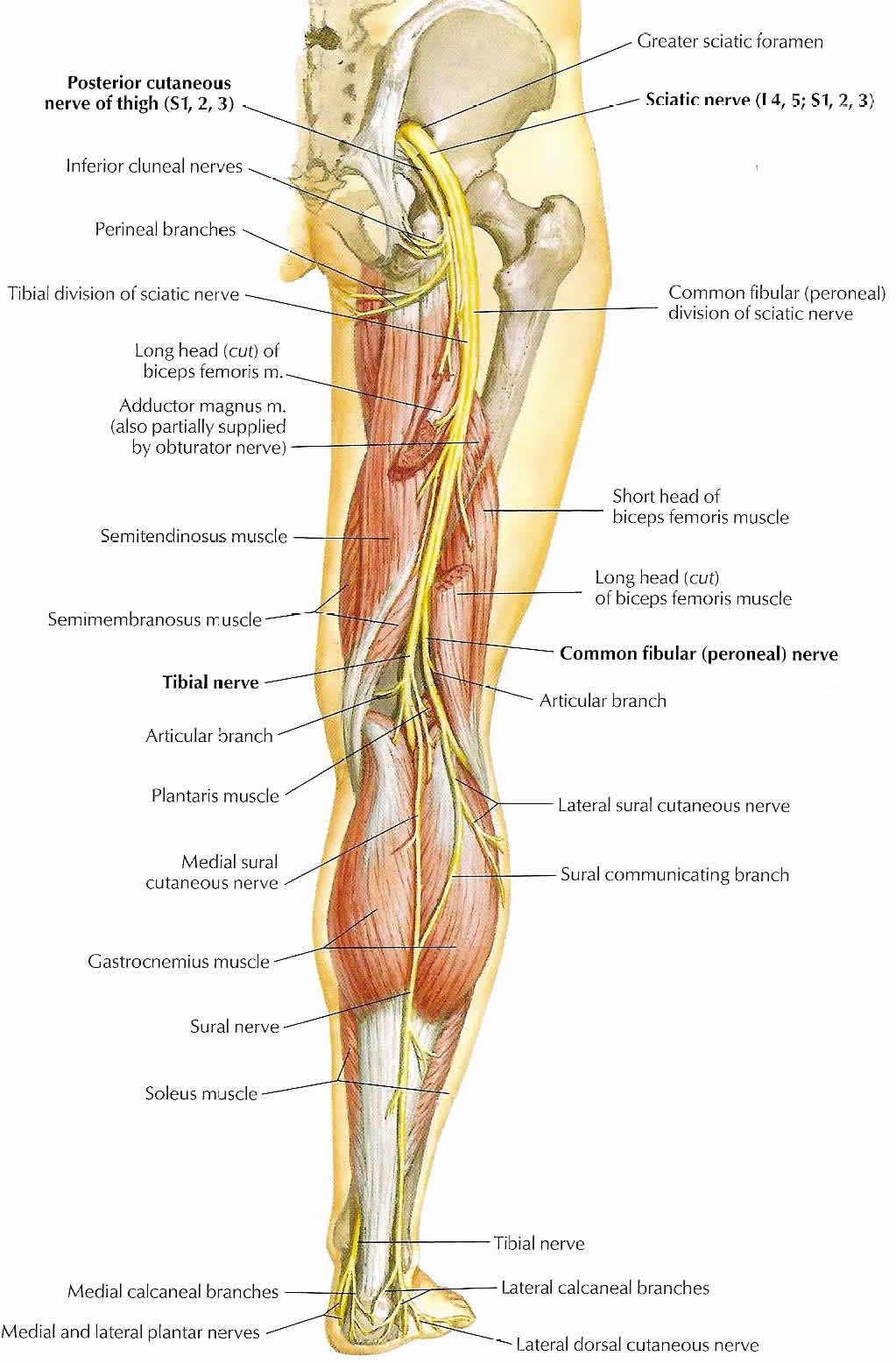
Also known as the ischiadic nerve or ischiatic nerve, the Sciatic Nerve is the largest nerve in the human body. The Sciatic Nerve runs down the leg behind the bicep femoris and powers the thigh muscles.By KDS4444 - Own work, CC BY-SA 4.0, https://commons.wikimedia.org/w/index.php?curid=53368293

The nerve begins in the Sacral Plexus Gray Sacral Plexus


as you can see from contrasting the above depictions of the nerve. Notice the outer thigh innervation and middle leg innervation from the upper nerves in the second photo. Contrast that to the inner thigh/back-leg innervation from the lower set of nerves. The sciatic nerve is a combination of the nervous tissue from L4 to S3 and continues down the leg to branch into the Tibial Nerve and the Common Peroneal Nerve at the popliteal fossa.

The Sacral Plexus and the Greater Sciatic Foramen

Here is a fantastic depiction of the sacral plexus and the nerve’s points of joining and separation through the Greater Sciatic Foramen which is covered by the piriformis. Here prentice-hall-sacral-plexusis a great view of the coccyx and sacral plexus which runs down the back of the leg. As the nerve travels, it is hammocked by the piriformis and then the bicep femoris before it branches. You can see a really great example of the support of the bicep femoris below

Posterior-View-of-the-Lower-Limb-Anatomical-Course-of-the-Sciatic-Nerve
Posterior-View-of-the-Lower-Limb


You can also see that as the nerve travels, it branches below the bicep femoris and the popliteal fossa which is also known as the knee pit. The biggest bone in the body, the femur supports and protects the sciatic nerve. We could definitely get into more detail about the branching of the nerve, but for now, let’s stick with the major components, we can get more specialized later.


Implications for your Yoga Practice

  1. If you haven’t started finding ways the stretch the muscles surrounding and supporting the biggest nerve in your body, its time to start. Finding ways to relax and stretch the piriformis and strengthen the sciatic nerve should be one of the primary goals of your practice. A healthy sciatic nerve will be most helpful in maintaining a pain-free leg!
  2. It is necessary to work into the layers of muscles surrounding the nerve tissue to truly release tension from it. This means that although an adjustment from a chiropractor might help in the short-term, you should be focused on re-aligning the leg muscles in your daily posture to create space for the sciatic nerve.
  3. Your hamstrings can be the primary instigator of your back pain! Quadriceps are filthy culprits as well! Find ways to stretch your legs and your back will often carry less tension as a result. And legs stretches can allow you to stretch the back in deeper ways. There are certain points inside of your hip/sacrum connection where your back and your legs are the same thing!
  4. This is a huge reason why downward dog feels so fantastic. You get to stretch the muscles around your biggest nerves! Downward can be one of the most sustainable yoga poses. It shouldn’t hurt! Just uncomfortable at first.
  5. Just to take the downward dog thing further, this is also why sun salutations are such a universal stretches in yoga and so good for relaxing the nervous system. I think sun salutations might be one of the best exercises you can do for your back.

15 Yoga Asanas for your Sciatic Nerve

  1. Hero’s Pose
  2. Downward Dog
  3. Foward Fold
  4. Sun Salutation A
  5. Low Lunge
  6. High Lunge
  7. Pyramid Pose
  8. Warrior 1
  9. Eagle Pose
  10. Triangle Pose
  11. Revolved Triangle Pose
  12. Half-moon
  13. Revolved Half-moon
  14. Half Pigeon
  15. Finishing Ashtanga Streches

References

Foot Reflexology Chart

Movement Shapes Your Body

Foot pain from your spine?

Your Foot Bone’s Connection to your spine

How can I break my Neck in my Foot


The Sciatic Nerve: A River of Energy Suppyling Human Legs Read More »

human bone anatomy

Human Bone Anatomy | Osteology

What are Bones?

Bones are not inanimate rock like structures in the human body; bones are organs that produce red and white blood cells, store minerals, enable mobility, and provide structural support for the body. They are lightweight, strong, and hard, and function within the body in many different processes, including autoimmune function. , There are two types of mineralized osseous tissue, or bone tissue, cortical and cancellous, and gives the bones rigidity and a coral-like three-dimensional internal structure. Other types of tissue found in bones include marrow, endosteum, periosteum, nerves, blood vessels and cartilage.

Primary Nutrients

Most literature proposes Calcium and Vitamin D as the primary nutrients for healthy bones.

Calcium is important in bone creation and repair. Your muscles, organs, and nerves also need calcium to function properly; nerves use sodium to pump electricity through nerves in the form of action potentials. Calcium helps to keep these actions potentials from excessively firing by working in concert with GABA receptors, most notably in high intensity auditory transduction. (http://phys.org/news/2007-03-calcium-life-death-nerve-cells.html). Leafy greens, fish, and some fruits are great sources of calcium.

Vitamin D is a group of secosteroids responsible for intestinal absorption of primary nutrients such as calcium, iron, and zinc. Vitamin D is synthesized in the skin is the primary way that the body produces the nutrient; though it acts as a hormone because the nutrient travels to become active in the liver and kidneys. Vitamin D has a significant role in calcium homeostasis (balancing) and production in the kidneys and liver. It also affects neuromuscular and immune function.

Protein, magnesium, Vitamin K, and phosphorus are also suggested as beneficial nutrients for bone health.

Bone Structure

bone_layer_image

Bone tissue, bone marrow, blood vessels, epithelium, and nerves make up the different types of bone cells. Tissue includes Osteoblasts and osteocytes, which are involved in the creation and mineralization of bone; osteoclasts reabsorb bone tissue. The mineralized matrix of bone tissue has an organic component of mainly collagen called ossein and an inorganic component of bone mineral made up of various salts. Bone tissue refers specifically to the bone mineral matrix that forms the rigid sections of the organ. There are two types of bones: cortical and cancellous. Cortical bone tissue create hard exteriors for protection while cancellous bone is more spongy and allows for the metabolic processes on the interior of the organ; the two are biologically identical, but the expression of their microstructures are specialized.

Bone marrow is flexible tissue and reproduces red and white blood cells as well as lymphocytes that support the immune system. Cores of marrow in the heads of long bones create about 500 billion red blood cells per day in hematopoiesis. 4% of human physiology is bone marrow; so about 5 pounds if you weight ~125. The body creates two types of marrow: red, the only type in the body at birth; and yellow, which increases in proportion during the aging process. Transplants can cure extreme diseases and is one of the primary reasons why stem cells can be so beneficial. The body stores marrow in the femur, hips, vertebrae, and ribs.

Osteo Factshttp://training.seer.cancer.gov/index.html

At birth, there over 270 bones in the body, which during the aging process turn into 206 by fusing together (joining). The biggest is the femur
(thigh) and the smallest is the stapes in the inner ear.  The hard cortical tissue (outer layer) comprises 80% of mass and networks of trabecular marrow comprise the rest. Bones are mineral reserves for the body and marrow stores fat. They are metabolically very active and work in tandem with the digestive system, immune system, and endocrine system in balancing nutrients, defending against disease, and releasing hormones, respectively. 22 bones fuse together after birth to form the skull. 26 aligned, specialized bones called vertebrae make up the spine, protect the spinal cord, and form the primary support structure for the body.

Aging and Osteoporosis

The problems arising from bones occur in osteoporosis, fractures, arthritis, tumors, and infections can affect the organic tissue. Fractures are breaks in tissue, from repetitive force or trauma. Aging causes osteoporosis; the body stops producing the necessary amount of building material for the body and literally means “holey bone” (porosis meaning hole). Tumors and malignancy’s can occur in various forms in bone tissue as well. This makes it much easier for the bones to fracture.

Cancer

Cancer can also occur in tissues structures and is a common site for it to metastisise to. Several primary cancers occur within the bones and some even within the marrow, such as Leukemia and multiple myeloma. The tissue distorted by cancer is normally more prone to fracture and weakness, which becomes particularly painful when it occurs in the spine.

References:

  1. AAOS – http://orthoinfo.aaos.org/topic.cfm?topic=A00317
  2. Wikipedia – https://en.wikipedia.org/wiki/Bone_marrow
  3. ASU Ask a Biologist – https://askabiologist.asu.edu/bone-anatomy
  4. Wikipedia – https://en.wikipedia.org/wiki/Neuroregeneration
  5. NOFG – https://www.nof.org/patients/what-is-osteoporosis/
  6. Skelton – http://www.innerbody.com/image/skelfov.html

Human Bone Anatomy | Osteology Read More »

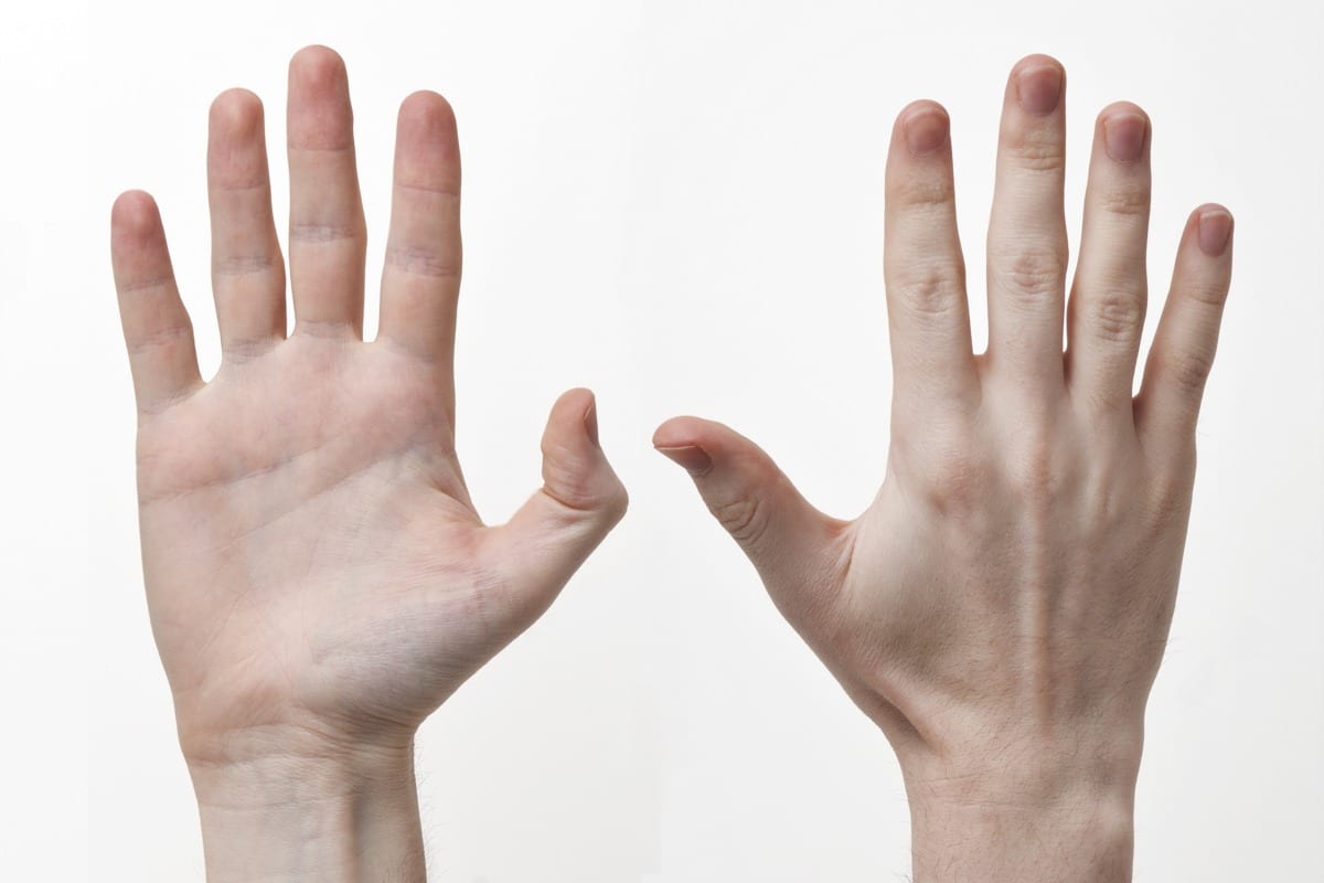
"Human-Hands-Front-Back" by Evan-Amos - Own work. Licensed under CC BY-SA 3.0 via Wikimedia Commons - http://commons.wikimedia.org/wiki/File:Human-Hands-Front-Back.jpg#/media/File:Human-Hands-Front-Back.jpg

Hand Anatomy, Physiology, and Use

The Usefulness of Man’s Hand

The hand is one of the most intricate and useful mechanisms of the entire human body; it is a prehensile (appendage for grasping) that humans share with chimpanzees, lemurs, and monkeys; even Koalas have opposable thumbs that are very similar to the thumbprints of the hands of humans. We humans absolutely have the ability to “think” with our hands; when we consider their connection to the brain we find the hand contributes to our thoughts and feelings. Fingers contain some of the most dense nerve endings on the entire body. The hand is greatest source for tactile feedback on the body and has the greatest impact on the sense of “touch”.

The hand has an intricate connection with the eyes and brain partially because they have the greatest mobility of any part of the human body. Each hand is paired with a dominant opposite side of the brain in the same fashion as the eyes. This “crisscrossing” of neuronal passageways occurs throughout the nervous system. The primary motor cortex is responsible for movement in the hands and body and executes movements in concert with the rest of the motor cortex.

There are 27 bones in the hand. 14 of which are in the fingers. There are 24 muscles groups innervated by various motor and sensory pathways that comprise 3 nerves: the radial, ulnar, and median nerves. These cascade to form 2500 nerve receptors per square centimeter on the surface of each hand.

Bones of the Human Hand

Lets start by looking at the bones. Each finger has three sections of bone: distal (fingertip), middle, and proximal; the thumb has two, theHand Bones middle bone is simply missing in between the top and bottom bones. The proximal bones connect to five metacarpals which connect to the eight carpal bones of the wrist. The fingers have 14 bones, the wrist has 13. The wrist has significantly more ligaments and less sensory nerves and mobility that the fingers. The bones of the hand_bones_detailed wrist are known as the carpal/carpus bones(from the Greek καρπὁς, “carp” means to pluck; an action the wrist performs) and there are eight of them (in order of ossification, or bone tissue growth): Capitate, Hamate, Triquetrum, Lunate, Trapezium, Trapezoid, Scaphoid, and Pisiform. Sometimes the radius and ulna bones are considered a part of the hand because of the role they play in the articulation of the wrist. There are also a large number of sesamoid bones in the hands (named after sesame seeds because they are so small). They are usually found near the thumb and are often formed in response to strain; they act like a pulley system for muscles and ligaments to slide over and spread muscular forces.

Ligaments and Tendons of the Hand and Wrist

In the hand, there are 18 ligaments that are separated into four groups:

  1. The ligaments of the wrist proper which unite the ulna and radius with the carpus: the ulnar and radial collateral ligaments; the palmar and dorsal radiocarpal ligaments; and the palmar ulnocarpal ligament.
  2. The ligaments of the intercarpal articulations which unite the "Braus 1921 201" by Braus, Hermann - Anatomie des Menschen: ein Lehrbuch für Studierende und Ärzte. Licensed under Public Domain via Wikimedia Commons - http://commons.wikimedia.org/wiki/File:Braus_1921_201.png#/media/File:Braus_1921_201.pngcarpal bones with one another: the radiate carpal ligament; the dorsal, palmar, and interosseous intercarpal ligaments; and the pisohamate ligament. (Shown in red in the figure.)
  3. The ligaments of the carpometacarpal articulations which unite the carpal bones with the metacarpal bones: the pisometacarpal ligament and the palmar and dorsal carpometacarpal ligaments. (Shown in green in the figure.)
  4. The ligaments of the intermetacarpal articulations which unite the metacarpal bones: the dorsal, interosseous, and palmar metacarpal ligaments. (Shown in yellow in the figure.)

In the image below, Hand_ligaments you can see how the blood vessels travel between the fingers next to the nerves and the padding of the hand on top of the ligaments used to keep the wrist bones compact as they rotate and move through space. The Ulnar nerve is on the left, near your pinky, and Grays_arm_nervesthe radial nerve is closer to your thumb and is almost entirely dedicated to its innervation and sensitivity. The median nerve is in the middle and acts as what is probably the primary sensory nerve. This nerve innervates your pointer finger and middle finger, which are your primary fingers for tactile sensing. There is a depiction from Gray’s anatomy on the right that shows how the three nerves flow through the arm down to the fingers.

The Hand’s Muscles Groups

I could probably write an article on each of the finger muscles exclusively. Bear with me as we go through these muscles groups. The muscles of the hand are some of the most sensitive and finely tuned muscles in the body. They are normally separated into two categories: extrinsic and intrinsic. Extrinsic muscles have their muscle belly (the majority of muscles fibers) on the forearm.

The intrinsic muscle groups are the thenar (thumb: Abductor pollicis brevis abductsFlexor pollicis brevisOpponens pollicis) and hypothenar (little finger) muscles; the interossei muscles originatingHand_muscles2 between the metacarpal bones; and the lumbrical muscles arising from the deep flexor digitorum profundus muscles (and are special because they have no bony origin) to insert on the dorsal extensor hood mechanism.

The fingers have two long flexors located on the underside of the forearm. The deep flexor attached to the distal phalanx (farthest) and the superficial flexor attaches to the middle phalanx. These are what allows your fingers to bend. The thumb also has two flexors, one long and one short and these work together with the thenar muscles to allow the thumb to grasp. The thumb is quite a complex mechanism in and of itself; kinda makes me want to write an article on it.

The extensors on the top of the forearm arrange in an even more complex way. The tendons unite with the lumbrical and interrossus muscles to form the extensorhood mechanism. The extensors straighten the digits. The thumb has two extensors on the forearm which form the anatomical snuff-box, or the triad at the base of your thumb. The pointer finger and little finger both have an extra extensor for pointing.

The Skin of the Hand

The skin of the hairless side of the hand (palm) is very thick and can be bent easily while maintaining connection with the muscles and bones of the hand. Palm skin is usually lighter because of inhibited melanin (skin pigment) production and therefore don’t tan. Fingerprints, or the papillary ridges exist to increase friction when the hand is grasping an object. The skin of the top of the hand is soft and pliable to allow the fingers to recoil quickly.

Conclusion

The hand is complicated, especially in terms of muscular innervation, but we are still learning enormous amounts about how they have evolved into their current state. Comparative physiology is very useful for this and we are constantly exploring more about ourselves through animals and our genetic ancestors. If you have any requests for articles, or interesting additions to this one, please ask. Feel free to add anything that I have missed, or to ask any questions in the comments.

sources (besides Wikipedia):
1. http://www.oandplibrary.org/al/pdf/1955_02_022.pdf (Craig L. Taylor PHD & Robert J Schwartz, MD)
2. http://www.aofas.org/footcaremd/conditions/ailments-of-the-big-toe/Pages/Sesamoiditis.aspx
3. https://ispub.com/IJFS/1/2/9047#sthash.lchtoImt.dpbs

Hand Anatomy, Physiology, and Use Read More »

hip_musculature_spinal_support

Anatomy of the Lower Back

(Part 1 of 2: Muscular Skeletal System)

If you have practiced yoga lately, chances are that you sat on the floor for a little while. This is a very healthy activity that every human should probably practice regularly for the strength of the pelvic floor muscles, and to allow the inner thighs and hips to relax. You can always work your way into it with blocks, props, cushions, pillows; you can do it while watching TV. It is good for releasing the muscles in the lower spine which have a strong connection with the hips, pelvic floor, abdominal muscles, lower back, and lower organs, including the sex organs and excretion organs; it’s good for all that important stuff.

In this article, I will speak specifically about the lower back and the anatomical features that you will want to be aware of as you practice yoga. If you have any of the following symptoms, you may want to begin a restorative yoga practice to assist in the alleviation of your pain, as well as begin to sit on the floor regularly. If it is too painful to start, contact a specialist or something like that, here are some symptoms of dysfunction:

  • Pain and stiffness in the back.
  • Pain in the buttocks and the legs, often in the back of the thigh.
  • Pain that worsens when bending, stretching, coughing, or sneezing.
  • Sciatic Nerve pain (pain in the hips, or back of your legs that shoots down the leg)

The lower back is really composed of three areas of the body: the lower spine, the hips and tailbone, and the abdomen. Since the spine is encircled by musculature, the abdomen, spinal muscles, and hips are all integral aspect of maintaining a healthy lower spine and therefore lower back.

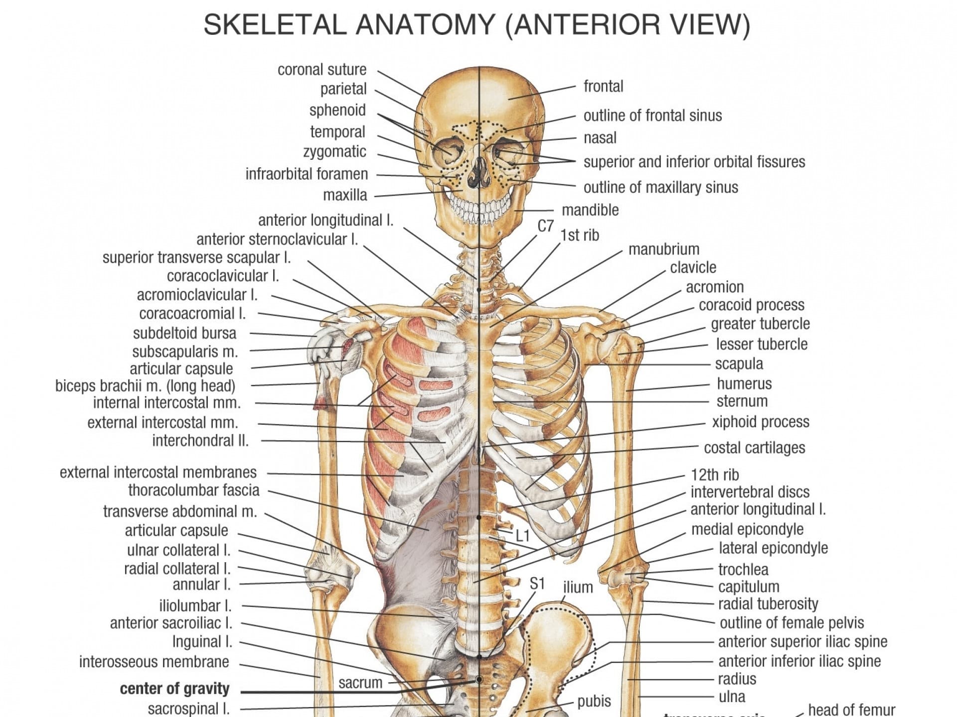
Here is a depiction of the skeletal frame with the lower back in red: lumbar_region from wikipedia

You can see the there is a lot of big bone support at the base of the spine you will know from your own body that your hips provide the support for the lower spine. The ribs and upper legs have a tremendous amount of connection with the lumbar region of the spine and are the primary support structures in providing space for the lower back and lowest organs. Here is a very detailed depictions of the inner hips muscles and lower spine: hip_musculature_spinal_support

This is a depiction of the primarily of the psoas muscles and illiacus muscles. The psoas is a primary muscle group that moves the trunk Gray's Quadratus Lombrumand is greatly affected by sitting habits. It lines the font of the spine and inserts separately into each vertebral process up to the T12 in most people. The illacus muscles line the insides of the hips and connect with the psoas at the insertion point of the lesser trochanter of the femur. Both the psoas and the QL run along the lumbar spine to the trunk, the QL going posterior to the spine and the psoas anterior to the spine, bone are connected to the transverse spinal processes. The muscles work together to move the trunk, along with the muscles of the abdomen. The psoas and the QL muscles are the primary muscles of the lower back, so we’ll come back to them.

There are a few more groups of muscles to pay attention to, but other primary muscle group to consider when talking about the lower back is the abdominals. Your abdominals provide frontal support for the spine, but in addition to the abdomen and primary lower back muscles of the Psoas and Quadratus Lumborum, the diaphragm, obliques, serratus muscles, pyramidalis muscles, levatores costarum, subcostal muscles, transverse thoracis muscles, and intercostal muscles play roles in the alignment of the lower spine. The final, possibly most under looked muscle is the latimus dorsi, which runs all the way along the back of the spine up to the shoulders. We can go over most of them as accessory breathing muscles, which is an action largely affected by the lower spine. It sounds like a lot of muscles because there are lots of muscles that are connected to your lower back. Let’s break it into pieces to see how it works.

Lets start at the top and work out way down the body, so lets start with the shoulders. The serratus muscles, obliques, levatores costarum, costal muscles and subcostal muscles all play a role in spinal alignment at the shoulder level. The subcostal muscles are the subcostal_muscles_ depictioninnermost, being inside of the rib cage, and surrounds the diaphragm along the ribs. The intercostal muscles are just superior, or further outside than the subcostal muscles. The levatores costarum run along the back of the spine on the outside of the rib cage, "Levatores costarum" by Uwe Gille - modified from Image:Gray389.png. Licensed under Public Domain via Wikimedia Commons - http://commons.wikimedia.org/wiki/File:Levatores_costarum.png#/media/File:Levatores_costarum.pngproviding even more support for the spine, which you can see act in opposition to the interlacing rib muscles. Notice the spinal erectors and spinalis muscle groups. You can also see how the muscles interweave with the spine and ribs, making breathing a full body movement. This is why forward folds are so effective at releasing the muscles the support the spine, so that they can stretch and relieve tension. This part of the reason why breathing in yoga can help to align the spine, and why spinal alignment and breathing have a close relationship. The obliques are a portion of the abdomen that you can read more about here. The serratus anterior is the another muscle to consider, which is also known as the punching muscle, as it pulls the shoulder blades forward. This is an extremely strong and useful muscle in yoga that supports you in handstands, forearm stands, and headstands in specific variations with proper alignments. and keeps the shoulders aligned, which then keeps the spine aligned. Like links on a chain. The final muscles to look at is theLatissimus_dorsi latimus dorsi, a muscle that runs from the lower back to the outside of the shoulder blades. The muscle connects the arms to the lower back, so can be really important for golfing, blowing, javelin throwing, or boxing. Anything where you are using your hips to power the upper body. These muscles can be easily overlooked in sun salutations, especially downward dog, which can allow the spine to hyperextend. This muscle is also more active in the elbow close push up, allowing the serratus anterior and lats to move the spine up from chaturanga into downward dog. Hollowing your armpits in plank/chaturanga/down-dog will likely activate and strengthen your lats, but its good to have a second pair of eyes on your alignment, so if you have questions find a local studio or teacher and ask them about your down dog. I’m sure they will be thrilled to answer your questions.

The last muscles to consider are the diaphragm, the obliques, the

Diaphragm
Diaphragm

pyramidalis muscles, and the transverse abdominus. The final piece of the puzzle is the rectus abdominus, which acts in direct opposition to the Psoas and QL muscles. The pyramidalis muscles are tiny triangles below the lowest layer of the rectus abdominus, and they form muscles just above the genitals. The diaphragm forms the inner musculature needed to move the ribs as the lungs expand.

abdomen image from http://www.usra.ca/The obliques line the outsides of the torso at the bottom of the rib cage, and all four layers of abdominals meet at the linea alba and run down to the pyramidalis muscles. You can see the lowest layer of the abdominals, the transverse abdominus, which acts as a kind of weight belt to support the lower spine when heavy lifting with the back, or squatting. It works with the psoas and QL to keep the trunk stable. The rectus abdominus acts in opposition to the QL and psoas, which forms a kind of push-pull system for you to lean forward and back, to squat, and to jump. Think of them as working against each other, but really they work in unison to support your spine. A great way to feel all of these muscles is to do burpees and/or sun salutations.

That wraps up the muscular and skeletal portions of the anatomy of the lower back. Please check back in about a week for the second section where I discuss nerves, organs, blood vessels, and fluid distribution, and if you are looking for something a bit more entertaining, you can check out the WANDERER series, I am working on part 13 right now and should have it out in a few days. Thanks for reading, would love to hear any questions or feedback


Anatomy of the Lower Back Read More »

The Respiratory System

Anatomy of Lungs and Respiration

artiphoria-prana-entering-the-body
The Anatomy of Breath – Elliot

There is one thing you have done every moment of your life. Even before you can remember. This will be the last thing you do before you die. And your awareness of this thing will partially determine how you exist on planet Earth. Prana, or your breath, is the primary mover on life in your body and corresponds deeply to your mental and physical health.

Humans have two lungs and five lobes, two on the left and three on the right (the right is bigger), each of which can be from 70-100 square meters in surface area, about the same surface area as a tennis court. The lungs have 2,400 kilometers of airways and 300 to 500 million alveoli which are gas exchange points for the bloodstream. These are powerful organs of exchange with the environment, with power and functioning that should not be taken for granted.

The respiratory system is a series of organs responsible for intaking oxygen from the atmosphere and expelling carbon dioxide back into the air. This basic gas exchange between the body and the atmosphere is completely dependent upon the respiratory system and almost every vertebrate animal has one. This exchange affects every other system, as they oxygenation of blood is necessary in every organ. The nervous system also seems to draw energy from the respiratory system, and the cardiovascular system takes cues from the respiratory system (both cue off brain activity) to determine how much blood it should be pumping based on breath rate. When the sympathetic nervous system becomes active (the flight or fight mechanism), heart rate is increased, respiratory rate is increased, the sensitivity of the nervous system is heightened to allow for survival, but this comes at a cost.

Yoga focuses primarily on the respiratory system’s functioning to move the muscular-skeletal system in the opposite way. In our modern world full of non-environmental stress and high levels of adrenaline in non-life threatening situations, the sympathetic nervous system is overactive and is probably the biggest contributor to the high fatality rates from cardiovascular disease (nutrition would be the other competing contributor). The respiratory system is vital to the functioning of every mammal on the planet and is one of the most intricate and powerful tools for surviving, prospering, and thriving on planet Earth.

LadyofHatsJmarchn – Own work using: Sobotta, Johannes (1982) Atlas der Anatomie des Menschen / 2 Brust, Bauch, Becken, untere Extremitäten, Haut. (18th ed.), Munich: Urban & Schwarzenberg ISBN3-541-02828-9OCLC260005032. Gray, Henry (1980) Gray’s Anatomy(36th ed.), Edinburgh: Churchill Livingstone ISBN0-443-01505-8OCLC7775214. Yokochi, Chihiro (1991) Atlas fotográfico de anatomía del cuerpo humano (3rd ed.), Mexico: Interamericana/McGraw-Hill ISBN968-25-1677-3OCLC33318149. Also used several online diagrams like:[1] [2]

I honestly think the vast majority of people take breathing for granted. Most Americans are in such a rush that they don’t even notice their superpower of consciousness. We don’t learn about breathing in school, or in early sports, which is really a shame because breathing concentration allow for intense amounts of focus. Every athlete should learn breath control techniques from young ages; I can remember when I learned to run with proper form at 15 and I think that learning about breathing should happen even younger. This is what keeps us all alive, after all, and we really should learn how to keep our nervous systems functioning optimally through breathing exercises.

To really understand how intricately related the nervous system and respiratory systems are, we need to go back in time to when you were born. At birth, a babies lungs are full of fluid, but once the child is released from the birth canal, the central nervous system trigger a huge change in reaction to the environment, which then triggers the first breath, about 10 seconds later. From there, the lungs develop rapidly until at about 2, the alveoli are fully developed, then the lungs begin to grow normally until full adult muscular maturity is reached. The lungs are muscular and most mammals use their musculoskeletal systems to support their breathing, as humans do. This is why yoga can alleviate many hampering disabilities having to do with lung functioning, because strengthening the accessory muscles to the diaphragm strengthens the overall functionality of the respiratory system.

The muscles of the respiratory system are the following:
  • the diaphragm (primary)
  • the external intercostals
  • the internal intercostals (intercostals interlace on the inside and outside of the ribs).
The accessory muscles are:
External-and-Internal-Intercostals-of-the-Thoracic-Cage

As you can see, there are a tremendous amount of accessory muscles involved in breathing. I interpret this a particular way, that there is an enormous spectrum between thriving and breathing with ease and freedom contrasted to breathing for survival, or breathing only with the diaphragm and ribs, which puts extreme amounts of stress on those muscles. I think the idea of balance between the primary and accessory muscles is the right idea, and the stronger the accessory muscles, the more powerful breathing will follow. This takes time, muscles build strength in increments, and this is probably the biggest reason why yoga is so difficult for many Americans. Because we need it the most!

Questions

  1. What kinds of breathing exercises do you practice for optimal health?
  2. What kinds of breathing exercises would you like to learn about?
  3. Do you find that breathing affects your mental health?
  4. Do you find time to meditate on your breathe during the day?

References

  1. Teach Me Anatomy – Thorax
  2. Wikipedia – Respiratory System

The Respiratory System Read More »

Scroll to Top

Subscribe

Get email updates of new articles when they are published

We promise we’ll never spam! Take a look at our Privacy Policy for more info.