Human Shoulder Anatomy
Understanding shoulder anatomy can help to avoid injury, promote rehabilitation, and can assist you in using the joint optimally. Please let me know if you have questions about this article in the comments section at the bottom of the page.
The human shoulder is a powerful and large anatomical structure. The hinging ball and socket joint allows for vast gains in momentum over short periods of time and is relatively versatile. The shoulder anatomy allows for many types of throwing, fine motor movement down to typing, powerful grasping, hefting objects, climbing, combat, quadruped movement, etc. The shoulder also has a large range of motion; however, this makes the shoulder prone to injury.
Bones of the Shoulder
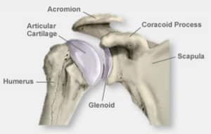
The shoulder joint is relatively loose. There are three main bones of the shoulder: the collarbone, shoulder-blade, and the upper arm bone. These are known as the clavicles, scapula, and humerus, respectively. The shoulder blade also has a bone called the caracoid process which connects to the biceps at the front of the arm and an upwardly angled bone called the acromion that connects to the Clavicle (collarbone) via the CA ligament.
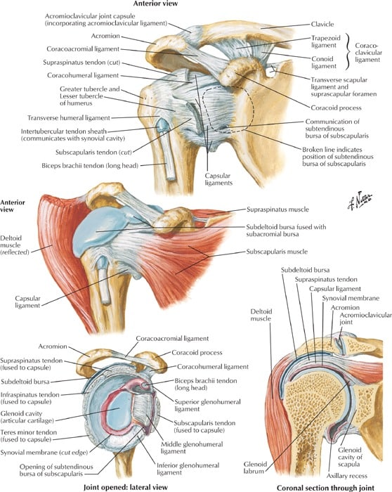
Ligaments of the Shoulder
There are large amounts of ligaments and tendons in the shoulder joint, because of its versatility, stability, and strength. As you can see, the three bones of the joint are combined together with vast arrays and webbings of ligaments that allow for the large range of motion while keep the joint stable. Honestly, the joint is so complex that using words to describe it become somewhat useless. So here’s a huge blown up picture for you to look at in awe of how fucking amazing your shoulders are:
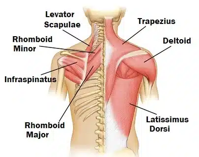
Muscles of the Shoulder

All of the deep ligaments that you see above are supported by muscles tissue. The muscles that make up the rotator cuff are the infraspinatus, supraspinatus, subscapularis, and teres. There are also three deltoid muscles on the head of the humerus, the rhomboids that connect the shoulder to the spine and the traps which connect the shoulder and neck, and provide support for the shoulder blades.
The infraspinatus muscle runs along the scapula (shoulder blade), covering the back of it over the teres minor muscle. The teres minor connects the outer arm with the outer lower edge of the shoulder blade. The supraspinatus connects the head of the humerus (arm bone) to the inside edge of the scapula articulating underneath the clavicle (collarbone). The teres major connects the outer clavicle with the back of the humerus; it is more superficial and larger than the teres minor. The subscapularis muscles run on the inside of the shoulder blade, but is not connected to the rib cage which is part of what allows the shoulder blade to have such a broad range of motion. Over all of these muscles are the deltoids, which are the most superficial shoulder muscles. They are separated into anterior, lateral, and posterior sections.
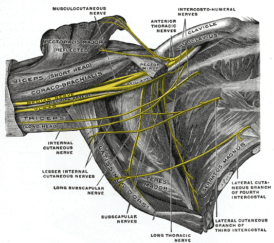
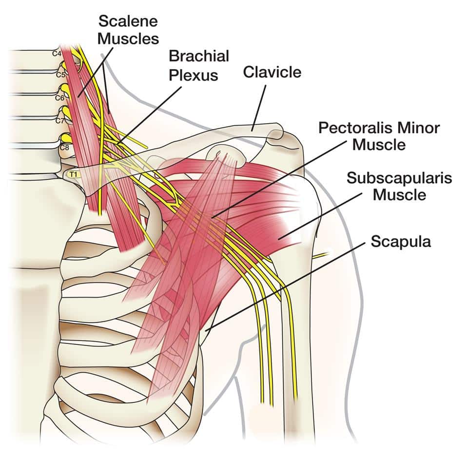
Nerves of the Shoulder
The nerves of the shoulder are also complex; consider that the fine motor function of typing must travel from your spine to your fingertips through the intricacies of the shoulder joint. You also have a very responsive feedback loop between your eyes and hands, which travels within the shoulder and into the forearms and fingers.
There are three primary nerves in the arm that run through the interior of the joint and connect to the digits (fingers).

The radial nerve provides innervation to the dorsal muscles of the arm: triceps, extrinsic extensors of the hands, as well as sensory innervation to the back of the hand, except for the pinky and half of the ring finger. It originates from the brachial plexus, carrying fibers from the ventral roots of spinal nerves C5, C6, C7, C8 & T1.
The ulnar nerve provides innervation to the back of the other three fingers, including the thumb. It also provides the majority of the innervation of the forearm and head of the bicep. The ulnar nerve originates from the C8–T1 nerve roots (and occasionally carries C7 fibers) which form part of the medial cord of the brachial plexus, and descends on the posteromedial aspect of the humerus.
The medial nerve provides innervation for the inside of the thumb, pointer, middle, and half of the ring finger. It also innervates the lateral and inferior portions of the forearm. The median nerve originates from the lateral and medial cords of the brachial plexus, and has contributions from ventral roots of C5-C7(lateral cord) and C8 & T1 (medial cord).
Here is a final picture of the brachial plexus to assist in visualizing how the nerves flow down the arms.
This concludes my article on shoulder anatomy; please write any questions below!