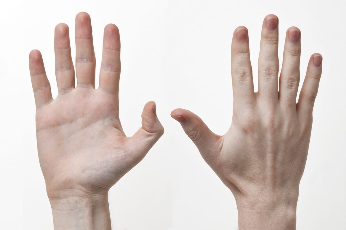
"Human-Hands-Front-Back" by Evan-Amos - Own work. Licensed under CC BY-SA 3.0 via Wikimedia Commons - http://commons.wikimedia.org/wiki/File:Human-Hands-Front-Back.jpg#/media/File:Human-Hands-Front-Back.jpg

Hand Anatomy, Physiology, and Use

The Usefulness of Man’s Hand

The hand is one of the most intricate and useful mechanisms of the entire human body; it is a prehensile (appendage for grasping) that humans share with chimpanzees, lemurs, and monkeys; even Koalas have opposable thumbs that are very similar to the thumbprints of the hands of humans. We humans absolutely have the ability to “think” with our hands; when we consider their connection to the brain we find the hand contributes to our thoughts and feelings. Fingers contain some of the most dense nerve endings on the entire body. The hand is greatest source for tactile feedback on the body and has the greatest impact on the sense of “touch”.

The hand has an intricate connection with the eyes and brain partially because they have the greatest mobility of any part of the human body. Each hand is paired with a dominant opposite side of the brain in the same fashion as the eyes. This “crisscrossing” of neuronal passageways occurs throughout the nervous system. The primary motor cortex is responsible for movement in the hands and body and executes movements in concert with the rest of the motor cortex.

There are 27 bones in the hand. 14 of which are in the fingers. There are 24 muscles groups innervated by various motor and sensory pathways that comprise 3 nerves: the radial, ulnar, and median nerves. These cascade to form 2500 nerve receptors per square centimeter on the surface of each hand.

Bones of the Human Hand

Lets start by looking at the bones. Each finger has three sections of bone: distal (fingertip), middle, and proximal; the thumb has two, theHand Bones middle bone is simply missing in between the top and bottom bones. The proximal bones connect to five metacarpals which connect to the eight carpal bones of the wrist. The fingers have 14 bones, the wrist has 13. The wrist has significantly more ligaments and less sensory nerves and mobility that the fingers. The bones of the hand_bones_detailed wrist are known as the carpal/carpus bones(from the Greek καρπὁς, “carp” means to pluck; an action the wrist performs) and there are eight of them (in order of ossification, or bone tissue growth): Capitate, Hamate, Triquetrum, Lunate, Trapezium, Trapezoid, Scaphoid, and Pisiform. Sometimes the radius and ulna bones are considered a part of the hand because of the role they play in the articulation of the wrist. There are also a large number of sesamoid bones in the hands (named after sesame seeds because they are so small). They are usually found near the thumb and are often formed in response to strain; they act like a pulley system for muscles and ligaments to slide over and spread muscular forces.

Ligaments and Tendons of the Hand and Wrist

In the hand, there are 18 ligaments that are separated into four groups:

  1. The ligaments of the wrist proper which unite the ulna and radius with the carpus: the ulnar and radial collateral ligaments; the palmar and dorsal radiocarpal ligaments; and the palmar ulnocarpal ligament.
  2. The ligaments of the intercarpal articulations which unite the "Braus 1921 201" by Braus, Hermann - Anatomie des Menschen: ein Lehrbuch für Studierende und Ärzte. Licensed under Public Domain via Wikimedia Commons - http://commons.wikimedia.org/wiki/File:Braus_1921_201.png#/media/File:Braus_1921_201.pngcarpal bones with one another: the radiate carpal ligament; the dorsal, palmar, and interosseous intercarpal ligaments; and the pisohamate ligament. (Shown in red in the figure.)
  3. The ligaments of the carpometacarpal articulations which unite the carpal bones with the metacarpal bones: the pisometacarpal ligament and the palmar and dorsal carpometacarpal ligaments. (Shown in green in the figure.)
  4. The ligaments of the intermetacarpal articulations which unite the metacarpal bones: the dorsal, interosseous, and palmar metacarpal ligaments. (Shown in yellow in the figure.)

In the image below, Hand_ligaments you can see how the blood vessels travel between the fingers next to the nerves and the padding of the hand on top of the ligaments used to keep the wrist bones compact as they rotate and move through space. The Ulnar nerve is on the left, near your pinky, and Grays_arm_nervesthe radial nerve is closer to your thumb and is almost entirely dedicated to its innervation and sensitivity. The median nerve is in the middle and acts as what is probably the primary sensory nerve. This nerve innervates your pointer finger and middle finger, which are your primary fingers for tactile sensing. There is a depiction from Gray’s anatomy on the right that shows how the three nerves flow through the arm down to the fingers.

The Hand’s Muscles Groups

I could probably write an article on each of the finger muscles exclusively. Bear with me as we go through these muscles groups. The muscles of the hand are some of the most sensitive and finely tuned muscles in the body. They are normally separated into two categories: extrinsic and intrinsic. Extrinsic muscles have their muscle belly (the majority of muscles fibers) on the forearm.

The intrinsic muscle groups are the thenar (thumb: Abductor pollicis brevis abductsFlexor pollicis brevisOpponens pollicis) and hypothenar (little finger) muscles; the interossei muscles originatingHand_muscles2 between the metacarpal bones; and the lumbrical muscles arising from the deep flexor digitorum profundus muscles (and are special because they have no bony origin) to insert on the dorsal extensor hood mechanism.

The fingers have two long flexors located on the underside of the forearm. The deep flexor attached to the distal phalanx (farthest) and the superficial flexor attaches to the middle phalanx. These are what allows your fingers to bend. The thumb also has two flexors, one long and one short and these work together with the thenar muscles to allow the thumb to grasp. The thumb is quite a complex mechanism in and of itself; kinda makes me want to write an article on it.

The extensors on the top of the forearm arrange in an even more complex way. The tendons unite with the lumbrical and interrossus muscles to form the extensorhood mechanism. The extensors straighten the digits. The thumb has two extensors on the forearm which form the anatomical snuff-box, or the triad at the base of your thumb. The pointer finger and little finger both have an extra extensor for pointing.

The Skin of the Hand

The skin of the hairless side of the hand (palm) is very thick and can be bent easily while maintaining connection with the muscles and bones of the hand. Palm skin is usually lighter because of inhibited melanin (skin pigment) production and therefore don’t tan. Fingerprints, or the papillary ridges exist to increase friction when the hand is grasping an object. The skin of the top of the hand is soft and pliable to allow the fingers to recoil quickly.

Conclusion

The hand is complicated, especially in terms of muscular innervation, but we are still learning enormous amounts about how they have evolved into their current state. Comparative physiology is very useful for this and we are constantly exploring more about ourselves through animals and our genetic ancestors. If you have any requests for articles, or interesting additions to this one, please ask. Feel free to add anything that I have missed, or to ask any questions in the comments.

sources (besides Wikipedia):
1. http://www.oandplibrary.org/al/pdf/1955_02_022.pdf (Craig L. Taylor PHD & Robert J Schwartz, MD)
2. http://www.aofas.org/footcaremd/conditions/ailments-of-the-big-toe/Pages/Sesamoiditis.aspx
3. https://ispub.com/IJFS/1/2/9047#sthash.lchtoImt.dpbs

Hand Anatomy, Physiology, and Use Read More »