grays_shoulder_anterior

Human Shoulder Anatomy and Physiology

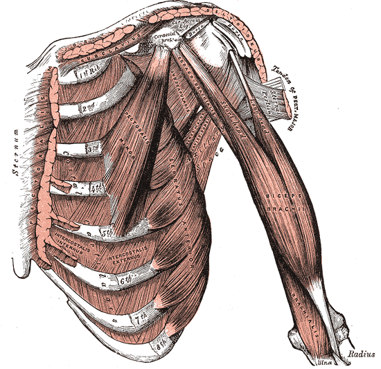
There are 3 bones in the human shoulder, or glenohumeral joint; the humerus, the clavicle, and the scapula. These bones are stabilized by 15+ muscles, depending on how you count them. These muscles function to stabilize the joint. This is what allows you to type, swing, and grasp with utter precision. Homo sapiens shoulder is precisely mobile, but lacks the stability and strength of our great ape cousins.

Gray's Shoulder Joint Depiction
Gray’s Anatomy

The muscles and bones of the shoulder joint work very closely together. They are very often depicted together in anatomy books because of how they functional in unison. The human shoulder joint is nothing short of incredible as a feat of natural evolution. It is a major evolutionary advantage over our primate cousins. Human beings the ability to climb, sprint, and perhaps most incredibly to throw objects accurately over large distances in conjunction with the excellent eye-sight of homo-sapiens sapiens because of our shoulders. And we can still climb, but must use our legs dominantly.

The Clavicle and Scapula are both considered to be part of the shoulder girdle, the structure that supports the appendages of the upper body. The shoulder provides stability for the neck, or upper third of the spine.

Bones of the Shoulder

Humerus

Scapula – wing bone, or blade bone connects the humerus and clavicle and lies on the back of the rib cage. The name derives from early Roman times when it was thought that the bone resembled a trowel or small shovel.

Humerus – the humerus is a long bone of the shoulder joint, connecting the shoulder girdle to the forearm.

Clavicle – also known as the ‘collarbone’, it is the first bone to ossify in an embryo, and connects the sternum to the scapula. It rotates upon its axis like a key when the shoulder is abducted. It is also the most commonly fractured bone.

Tendons and Ligaments of the Shoulder and Armpit

Gray's Shoulder Tendons

The Glenoid cavity is a shallow depression in the scapula, that connects to the head of the humerus and allows for the arm-bone’s articulation, forms the basis for the ball and socket joint and is held in place by the head of the biceps tendon. The rotator cuff also reinforces this joint with the supraspinatus tendon.

The Rotator Cuff consists of four primary tendons: the supraspinatus muscle, the infraspinatus muscle, the teres minor, and the subscapularis muscle. The tendons of these fours muscles merge to form the rotator cuff tendon.

The Coracoacromial ligament connects the coracoid process (the hook like structure on the shoulder blade) and the acromion (the highest profusion of the shoulder blade). This ligaments helps to shield the head of the humerus.

Gray's Rotator Cuff Ligaments

The AC Joint, or Acromioclavicular joint is the joint at the top of the shoulder that connects the acromion to the the collar-bones. There are several acromioclavicular ligaments as you can see in the image on the right and they are organized to provide added stability to the joint and to house the bursa and synovial fluid that allows the joint to articulate easily.

The conoid ligament connects the clavicle and the coracoid process further stabilizing the collar bone to the shoulder blade.

The caracohumeral ligament connects the coracoid process to the humerus.

Together, these ligaments stabilize and support the shoulder joint, allowing for the extreme mobility that we humans enjoy. However, the large amount of smaller ligaments and tendons sacrifice a certain amount of stability for this increased mobility and range of motion.

Shoulder Muscles

  1. Deltoid – responsible for lifting the arm and giving the shoulder its range of motion. Often this muscle is separated into 3 sub-muscles, anterior, lateral, and posterior as they are able to innervate separately.
  2. Teres Major – A small muscle that runs along the lateral border of the scapula and connect to the humerus.
  3. Teres Minor – extends laterally and obliquely from the head of the humerus to the scapula, underneath the Teres Major. This rotator cuff muscles rotates the head of the humerus and stabilized it as it moves in space.
  4. Supraspinatus – connects the scapula to the humerus and abducts the shoulder and arm.
  5. Infraspinatus – connects from the medial side of the scapula to the humerus to aid in stabilizing the shoulder. A thick layer of muscle on the outside of the shoulder blade and is the main external rotator of the shoulder.
  6. Subscapularis – Directly opposes the infraspinatus muscle on the interior of the shoulder blade. It rotates the humerus medially and adducts it, preventing the displacement of the humerus during motion.
  7. Serratus Anterior – originates on ribs one through eight and connects to the medial interior edge of the scapula. The serratus anterior muscles work in conjunction with the latissimus dorsi to lift the shoulder blades and pull them forward and are one of the primary core support structures for the shoulder. Shoulder injuries often occur in yoga because this muscle is not fully contracted, especially in Chaturanga.
  8. Subclavicus – A small muscles that lies between the clavicle and the first rib that draws the shoulders down and forward.
  9. Pectoralis Minor – a thin and flat muscle in the upper torso that lies underneath the pectorals major and originates in the second, third, and fourth ribs. (sometimes the 5th rib instead of the 4th). This is the primary chest muscle that assists in lifting the shoulders.
  10. Sternocleidomastoid – the primary visible neck muscle that rotates and turns the head and neck. It inserts at the sternum and clavicle and travels up to the mastoid at the temporal lobe of the skull.
  11. Levator Scapulae – the main function of this muscle is to lift the scapula, originates in the neck C1-C4 and travels down to the medial border of the scapula. Works in a state of near unison with the serratus anterior muscles.
  12. Rhomboid Major – connects the shoulder blade to T2-T5 of the mid spine. It is slightly deeper than the trapezius and slightly inferior to the rhomboid minor. Together with the serratus anterior and pectorals minor, it connects the shoulder blades to the rib cage.
  13. Rhomboid Minor – Also connects scapula to the spinal vertebrae, but superior (higher) than the rhomboid major and slightly smaller. Connects C7 and T1 to the shoulder blades. Oftentimes this muscle is completely fused with the Rhomboid major.
  14. Trapezius – a large paired surface muscle in the shape of a diamond, connecting the occipital lobe to the shoulder blades and travels down to the lower thoracic vertebrae. It helps to move the scapula and the arm. Because it connects both the spine and the shoulder blades, this muscle can be one of the primary causes of neck tension in the body.
  15. Latissimus Dorsi – a large flat muscle one the back that originates in the mid and lower back and travels all the way up to the head of the humerus. Is it the largest muscle in the upper body and is implicated for cardiac support and is also an accessory breathing muscle. Tightness in this muscle has been shown to be a primary contributor to back pain.
Gray's Brachial Plexus

Nerves of the Shoulder Joint

The Brachial Plexus is a network of nerve tissue that supplies the arm and shoulder with innervation. Branches of the plexus, in particular from C5-C6, supply the majority of the muscles of the shoulder. The plexus continues down the arm to form the radial, ulnar, and median nerves of the arm.

Blood Vessels of the Shoulder

The blood Vessels of the shoulder function very similarly to the nerves (often in the body, nerves and blood vessels run in parallel to make the innervation of the muscle tissue more accessible to the nervous system. The Auxiliary artery becomes the brachial artery at the upper arm and continues down the arm to become the radial and ulnar arteries. Most of the blood vessels of the shoulder branch off the auxiliary artery.

Rotation in the Shoulder

BursaShoulder bursitis is a common cause of shoulder pain and occurs when the rotator cuff tendons are impinged, or unable to articulate properly. The shoulder bursa is extremely important as it creates smooth range of motion for the arm and shoulder to travel.

Rotator Cuff – the rotator cuff tears are another common cause of shoulder pain, usually cause by a tear in the supraspinatus muscle.

Range of Motion – As I discussed earlier, the shoulder’s range of motion is largely allowed for by the tremendous amount of ligaments, tendons, and muscles that work together to mobilize the arm. This comes at the sacrifice of stability. The stability of the shoulder comes from the muscle tissue, which can limit the range of motion in the shoulder, which may be healthy for the skeleton, especially under large amount of duress. It is easy to see this limited range of motion in body builders, whose muscles have gotten large enough to impede the motion of the shoulder. An appropriate balance between stability and flexibility is what we are looking for in yoga (or at least I am looking for this balance) so that the joint can have maximum longevity.

All Referencing from Wikipedia.org

Images

Gray’s Book of Anatomy

Encyclopedia Britannica

Human Shoulder Anatomy and Physiology Read More »