yoga anatomy

leg_lateral_musculature

Keeping Knees safe in Hip Openers

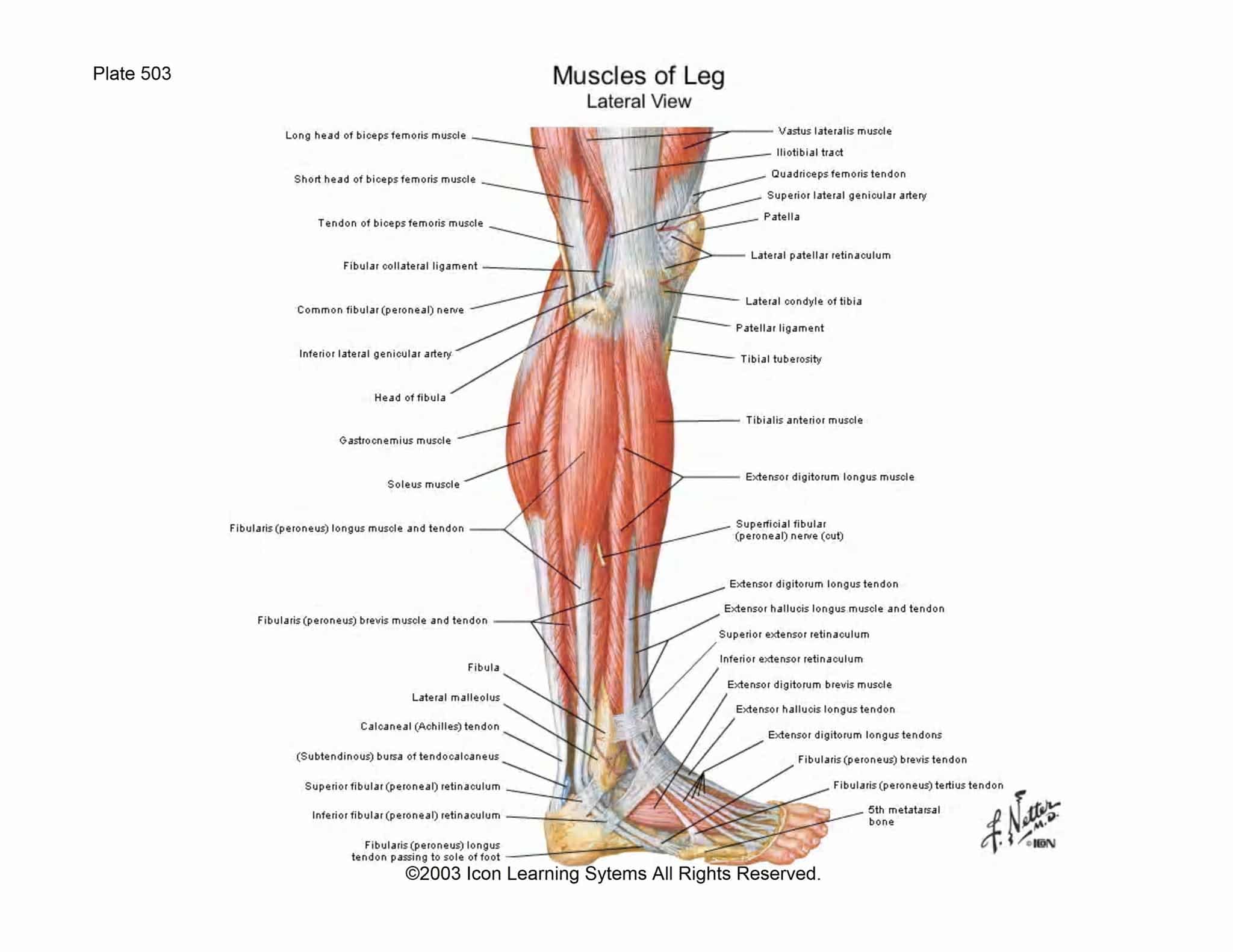
Over the past few months, I have been working diligently toward lotus pose. This has led me to some very stark realizations about how the knees need to be protected while opening the hips, at least for those of us with tight hips. This is an anatomy article geared at learning how to appropriately safely stretch with the ligaments of the knees, and how the opening of your hips is largely affected by your feet.

anterior_leg
anterior_leg

We can start with an overview of the anatomy of the outside of the leg. Lets start at the bottom and work our way up.

lateral_ankle_anatomy

There are a few major tendons in the ankle that need to be protected while opening the hip. If these tendons are not flexed during the stretching of the hips, stress will accumulate in the lateral portion of the knee, specifically in the lateral meniscus and the lateral collateral ligament.

Now we know the basic ligaments of the ankle and the outside of the knee that are being affected. Now let’s talk about the fibula, one of the most slender bones in the body.

lateral_ankle_ligaments

There is a tremendous amount of connective ligamentation on the outsides of the ankles, especially back towards the calcaneus where there are three ligaments to stabilize the ankle on the heel. The heel is a crucial point of stress when the hip is being opened and right on the lateral portion, or outside of the heel is where the most important flexion lies for the opening of the hips. This activation, which should continue up through the plantar and dorsal ligaments to the pinky toe of the foot. This will keep the ankle safe and properly aligned to allow the hip to open while the knee opens laterally.

Once the fibula is aligned properly, the outer knee ligaments and inner cushions can do their job and keep the interior knee ligaments from over stretching. You can see a great view of the knee’s mixture of interior and exterior ligamentation, which in reality overlap and interweave.

http://classroom.sdmesa.edu/eschmid/F07.11.L.150.jpg
knee ligamentation

The fibular collateral ligament is the most important ligament on the outside of the knee and is a thin sheath. This is the ligament that you really want to keep safe and relaxed while stretching the hip, which will ensure that the meniscus is not over-stressed. If it is, the ACL and PCL, the interior crossing ligaments of the knee could be overextended. Also keep in mind the moving the stretch further back into the hip will take time, so be patient with your bodies process. After all, yoga is all process, there is no completed pose or perfect posture. There are always possible improvements, different variations, alternate alignments.

Let’s move up into the thigh, and where you should be feeling the stretch. The IT band is a completely necessary activation, but it is really controlled by the flexion of the outer portion of the foot.

posterior_leg
posterior_leg


You can see here the muscles that should be receiving the stretch while the hips are externally rotated. The gluteus minimus, piriformis, gemellus muscles, obturator muscles, adductors, and quadratus femoris muscles. The medial gluteus also twists and stretches fairly significantly. There are also a lot of muscles at the front of the thighs that are receiving significant stress from the hip opening postures.

The Satorius muscle, Vastus Lateralis, and Tensor Fascia Latae stabilize the outsides of the knee. The pectineus and adductor longus muscles, adductor magnus, and gracilis muscles.

anterior_thigh_muscles

Armed with this information, you can now see how the foot and ankle have intricate connections all the way up the leg. Keeping the knee stable while opening the hip will allow you to practice more often, with less stress on our knees, something that I think we could all use a little bit less of. In addition, this article should allow you to know how the muscles that open your hip are stretched during external rotation. Any questions are greatly appreciated.

Keeping Knees safe in Hip Openers Read More »

Anatomy of the Rib Cage

The Rib cage is a primarily protective structure, encircling the heart and lungs. In your human body, normally you have (yes, if you can read this, you are human) 12 thoracic vertebrae connected to 24 ribs. The rib cage is also known as the Thoracic cage and is a core section of the human skeleton, provide support for neck, thorax, upper abdomen, and back. The word costal comes from Latin, where costae means rib.

The 7 highest pairs of ribs are known as the true ribs, because they attach to the sternum, the T shaped bone at the front of the chest. The 5 lower pairs of ribs, are known as false ribs because they connect to the costal (rib) cartilages above them. 2 of these ribs are known as floating ribs, because they do not attach at the front, and connect only to the lower thoracic vertebrae. The 5 lower ribs are what allows for respiration as the cartilage expandeds. The upper ribs are more functionally protective and attach to the sternum, the armored frontal plate for the heart and lungs.

The sternum has 3 primary parts:

  1. The Xyphoid process at the bottom – cartilage that normally becomes bone as an adult, is located at the same level as T6 and attached to the 7th rib and remaining lower ribs through their attachments to the 7th rib.
  2. The Gladiolus in the middle – this is the primary structure of the sternum, is flat, and is attached to the pec majors. It attaches to ribs 2 through 6.
  3. The Manubrium at the top of the rib cage – This articulates (moves) with the clavicles, or collar bones. It forms the base of the jugular.

At the floor of the Thoracic cavity the diaphragm expands and contracts and separates the lungs from the abdominals and organs. The top of the rib cage connects directly to the neck through the scalene muscles, and SCM. The serratus runs along ribs 1-8 and is the muscular outline of the ribs and connects the latimus dorsi and shoulder blades to the pectoralis major. Underneath the serratus you have the intercostals, which is a group of muscles intertwining in the ribs. All of these muscles play a role in breathing, if only for structural support of the rib cage and diaphragm.

Overall, the rib cage is one of the more elegant and multifunctional support structure for the human skeleton, musculature, and the major organs of the chest. See some more pictures below:

sternum anatomy
Torso Organs

Anatomy of the Rib Cage Read More »

Scroll to Top

Subscribe

ElliotTelford.com

We promise we’ll never spam! Take a look at our Privacy Policy for more info.