anatomy

Anatomy of your Tongue

The tongue is a muscular hydrostat (hydraulically powered food grabber in the same class as an elephant trunk, snake tongue, or octopus arms) with no support that acts as the organ for taste, or gustation. It lies the floors of the mouth of vertebrates and moves to manipulate nutrition for digestion and mastication (chewing).  It maintains constant pressure and is made of three directions of muscles and blood vessels to supply nerves and blood vessels. Many cultures also use the tongue phonetically, for specific communication (whistling, growling, kissing), or for cleaning the teeth and mouth.

There is a significant amount of musculature connecting the tongue tongue_musclesto the mouth. There are eight muscles in the tongue region, classified into intrinsic or extrinsic. The four intrinsic muscles change the shape of the tongue and are unattached to bone, while the four extrinsic muscles change the position of the tongue and are anchored to bone.

The extrinsic muscles are the hyoglossus, genioglossus, styloglossus,Gray_tongue_intrinsic palatoglossus that allow the tongue to extend outwards, retract, and move side to side. The intrinsic muscles of the tongue all originate and insert within the tongue. These muscles shape the tongue by lengthening and shortening, curling and uncurling, and flattening and rounding the surface. These muscles facilitate speech, swallowing, eating, and provides for the shape of the tongue. The average length of the tongue is about 10cm.tongue_arteries

The tongue receives blood through the lingual arteries, all of which drain into the internal jugular vein. The tongue is innervated by several nerves which carry the sensation of taste to the brain. The chorda tympani, the lingual nerve, the trigeminal nerve, and the glossopharyngeal carry the nervous information to the brain. The chorda tympani is particularly interesting, because it also innervates the muscles of the face, meaning there is likely a strong connection between facial expression and the sensation of taste and similarly, the trigeminal nerve is responsible for sensation in the face and the motor functions of biting and chewing. Together, these nerves create the highly specific feedback loop that ends up as the sensation of taste in the brain.

The tongue is covered with numerous taste buds, however, the sensations of different tastes are not localized to specific areas of the tongue. This was disproven and all taste sensations come from different parts of the tongue, though certain regions can be more sensitive to certain flavors. The different taste buds are filiform papillae, fungiform papillae, vallate papillae, and foliate papillae.

The taste receptors function by waiting for stimulus chemical to interpret, called tastants. Once a tastant has dissolved in saliva, it makes contact with the plasma membrane of the gustatory hairs, which are the site of transduction (conversion of one stimuli to integrate into the nervous system). The tongue is equipped with mostly taste buds on its dorsal (upward facing) surface, to sense the five different kinds of taste: umami, sweet, sour, bitter, and salty. Umami is currently the most researched and debated of the five kinds of taste.

Fungiform papillae, vallate papillae, and foliate papillae are the most associated with taste, while the filiform papillae is far more associated with increasing surface area of the tongue and to increase the friction between the tongue and food.

Bacteria builds up easily on the tongue and is the second most vulnerable soft tissue to pathogens, next to the gums. Tongue scraping can assist with removing debris and bacteria from the surface of the tongue. This can also be done with a brush, but I think that both are extremely useful for keeping the oral cavity clear of pathogens and potential disease. Most vertebrate animals have and use tongues, some are specifically adapted to catching prey, or to clean and groom fur, clear nostrils, or to regulate heat in the case of a dog. The tongue is an organ that has evolved over a long period of time and is extremely useful for animals that live above the sea-level.

That does it for the tongue, this will lead into the final bandha article, Jihva bandha, so check back soon to see more details about how to use the tongue while practicing. Talk to you soon!

Anatomy of your Tongue Read More »

Endocrine System – The Body’s Way of Talking

Endocrine System: Chemical Communication

The endocrine system refers to a collection of glands that secrete hormones into the circulatory system to target a distant organ with chemical messages. These tend to be slower processes, such as growth, menstrual cycles, or circadian rhythms, but also refer to procedures for dealing with stress and the environment. The notable endocrine glands are:
  • the pituitary gland – about the size of a pea, this gland protrudes atpituitary gland - the Endocrine System the base of the hypothalamus, there are two lobes in humans that regulate growth, blood pressure, pregnancy and childbirth, breast milk production, sex organ functioning, thyroid gland function, metabolism, osmolarity and water balance, temperature regulation, pain relief and sleeping patterns (but this is mostly the responsibility of the pineal gland. This is the seat of control of the mind over the entire body, hormonally speaking.
  • the pineal gland – The gland produces melatonin to regulate sleep cycles and circadian rhythm and the hormone is very sensitive to light. Descartes viewed the gland as the gateway between the body, mind, and soul and considered it to be the third eye. When children become teenagers, production becomes delayed leading to later sleeping and waking times.
  • the pancreas – largely regulates the digestive and lymphatic pancreas - the Endocrine Systemsystems, secreting insulin, glucagon, somatostatin, pancreatic peptide, and assists in digestion by secreting pancreatic which is a blend of digestive enzymes
  • ovaries, testes – ovaries release: estrogen, testosterone, progesterone, and unfertilized eggs; testicles release sperm and androgen (steroid hormones, primarily testosterone)
  • thyroid gland – one of the largest endocrine glands with two lobes around the trachea, below the thyroid cartilage also known as the Adam’s apple. It controls how quickly the body uses energy, makes proteins, and uses other hormones. It does this by producing hormones that regulate growth and rate of function of othethyroid gland - the Endocrine Systemr systems. Hormonal output of the thyroid gland is controlled by thyroid stimulating hormone produced by the anterior pituitary lobe, which itself is regulated by thyrotropin-releasing hormone produced by the hypothalamus
  • parathyroid gland – Humans usually have four, located on the back of the thyroid gland that produce parathyroid hormone and calcitonin. Together, these hormones regulate bone physiology including calcium levels and phosphate levels and reabsorption levels via the kidneys, so that the levels of these chemicals are optimal for the nervous and muscular systems to function
  • hypothalamus – a small portion of the brain located just above the brain stem with specialized cells to link the nervous system to the endocrine system via the pituitary gland. All vertebrate brains have a hypothalamus, which produce neurohormones that stimulate the secretion of pituitary hormones, controlling body temperature, hunger, thirst, and fatigue, different processes of the autonomic nervous system.
  • gastrointestinal tract – releases hormones to help regulate the digestive process, including gastrin, secretin, cholecystokinin, and ghrelin
  • adrenal glands – endocrine glands that sit atop the kidneys, responsible for stress hormones cortisol and corticosteroids, and also catacholamines, like adrenaline and noradrenaline. These cells also produce androgens to assist the body in dealing with stress, and the glands activate both the nervous and endocrine systems to work react to stress.

These glandular organs together make up the endocrine system, which sends chemical messages throughout the body, similar to the nervous system, however, the effects and mechanisms of the system are very different in their processes.

Glands? What are those?

The word gland is used for any organ that is used to excrete hormones for release into the bloodstream or into cavities inside of the body or to its outer surface. There are also two types of glands to be aware of: endocrine and exocrine; the latter uses ducts and bodily cavities for transport while the former uses the circulatory system for transport.

The exocrine system is a collection of glands that operates differently than the endocrine system, but is extremely useful to the body. The exocrine system uses three different methods of excretion:

  1. Merocrine glands – cells excrete their substances by exocytosis (cellular waste removal by containing the waste,then releasing it
a mammary gland
a mammary gland – the Endocrine System

outside of the cell): i.e. pancreatic acinar cells

  • Apocrine glands – cells that excrete by concentrating their waste, then cutting of a bud of the cell with the waste
  • Holocrine glands – The entire cell disintegrates to release its substance
  • It is easiest to understand the exocrine system by how it functions: it comprises sweat glands, salivary glands, mammary glands, and the liver. Together, the exocrine and endocrine systems work all of the glands of the body to remove waste.

    Endocrine System Bodily Operations

    The endocrine system operates differently from the exocrine system, in that it works via the circulatory system. Many organs have secondary endocrine functions in addition to their primary roles in the body. Endocrine organs usually signal themselves in groups and in certain orders, referred to as an axis. The Greek words ἐνδο- endo-, inside or interior and κρίνειν krinein, to separate or distinguish are  useful for understanding what the endocrine system does. It is essentially specializing specific organs to work in unison for specific bodily functions, including growing, sleeping, and the most basic of physiological needs, such as hunger and urination. In unison with the nervous system, this is how the body communicates internally.

    I will go into more detail in later articles about the particular neuroreceptors and hormones that the body uses for specific functions. Keep in mind that many are still unclassified and have properties that have not yet been observed and measured. The science of endocrinology and neurology have vast horizons to cover before we truly understand how these systems interrelate and function to produce conscious thought. But here are some great graphics of the known hormones and their organs of origin, thanks wikipedia!

    Endocrine_central_nervous


    Endocrine System – The Body’s Way of Talking Read More »

    Jalandhara Bandha | जालन्धर बंध (Bandhas part 3/4)

    Jalandhara bandha is an interlock tucking chin into chest to elongate the back of the neck. This is particularly useful in inversions like headstand and shoulder stand, which can place stress on the neck if it is not properly elongated. This interlock can also include opening the chest and sternum in many cases to allow for great lung expansion when inhaling.

    The sanskrit meaning of the word is enlightening: Jala means web, or net and dhara means contracting. So the contraction of the chin towards the chest lengthens the posterior (rear) neck muscles to elongate the back of the neck and highest portion of the spine. The neck muscles truly are an interweaving web to allow for the massive amount of rotation and movement that our heads are capable of. neck_muscles_detail

    By tucking the chin to chest, you allow the spine to grow longer, creating more room for breath neck_veins_detailcapacity (the spine is intricately linked to respiration). This is why the jalandhara bandha is used often in breathing exercises. Lengthening the rear neck muscles also creates more space for blood flow and nerve connections to the brain and skull. The arteries and veins that run along the neck muscles are extremely important; they transport oxygenated blood to the brain. This is why headstands and shoulderstands are so beneficial; they reverse this bloodflow and while the jalandhara bandha is locked allows for the nervous system to reverse it’s usually flow against gravity. The lymph system also receives enormous benefit from being inverted for an appropriate period of time.

    There are also some essential organs that are compressed during the interlocks. The thyroid gland get compressed during the lock, which can create more space for functioning of the organ. The lymph system is also greatly affected by the interlock, because compressing the lymphs will also create more room for the flow of lymphatic fluid. The physical benefits of the interlock are undeniable.

    neck_fullanatomy_details neck_greys_anatomy

    You can see the muscles of the neck in more detail and how the interlock is truly a contraction of a web of muscles surrounding the Hyoid bone. We’ll talk more about the hyoid bone in the next article, part 4: Jiva bandha. You will also notice a release in the shoulders when practicing the interlock, because of the relationship between the neck and the shoulders. You can see the trapezius muscle extends all the way to the back of the skull and that lengthening this in combination with the serno-cleido-mastoideus muscle. Muscularly, the contraction is extremely important for headstands! You should not be practicing balancing on your head without this interlock! It will lead to neck pain and possible injury. Headstand, according the yoga alliance statistics, is the pose where people get injured the most often in the United States. This is probably a result of not knosasankasanawing how to fully extend the neck using the jalandhara bandha interlock. If you want to start learning about the lock without a chance of injuring yourself, start in shoulderstand. Lengthen your neck as much as possible and then practice building the strength necessary for headstand in rabbit pose (sasankasana).

    Here is Leslie Karminoff’s depiction of shoulderstand, this is a great way to work on the bandha. I highly recommend checking out his work at Bandha Yoga.neck_muscles

    Compress chin to chest during meditation at the end of exhales. You can retain the lock for an inhale as well, notice the increased space on your inhale. Combine this with the uddiyana bandha and mula bandha and then notice how much space there is. This is a full lengthening of the spine through muscular contractions and muscular interlocks, which is extremely useful for creating space for breath, and life force energy.

    Stay tuned for the final piece of the puzzle of the bandhas, the Jiva bandha. Used in conjunction, these interlocks will change the way you practice. Stay tuned for part 4, talk to you soon…

    Jalandhara Bandha | जालन्धर बंध (Bandhas part 3/4) Read More »

    Transversus_abdominis

    The Anatomy of the Abdomen (part 3/4: Transversus Abdominis)

    Part I
    Part II

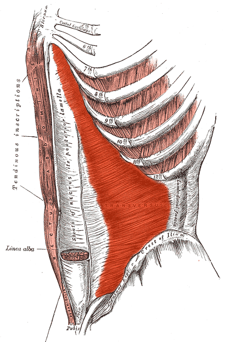
    The Transversus Abdominis is the deepest layer of the abdominal wall, underneath the internal obliques. It stabilizes the mid-back and pelvic areas. Since it is the deepest layer of abdominal tissue, its function is important is activating the other muscles of the abdomen and stabilizers in the spine during dynamic movement. It is also implicated in most spinal injuries though the scientific community is in debate about how much effect the muscle has on lower back dysfunction.

    The muscle originates in the borderline of the ribs, the front of the hip plate and the inguinal ligament, and the lumbar fascia. It joins the rectus sheath (6 pack muscles outlined in the abdomen) at the front of the abdomen and inserts in the linea alba. It joins at the front of the pubic bone via the conjoint tendon, which often conjoins with the internal oblique, but can also be separated.

    The transversus abdominus muscles is also known as the corset transverse_abdominismuscle, because it holds in the organs and abdominals. It is innervated, or activated, with T7-T11 and nerves from the hip plate
    and is extremely useful in stabilizing the spine. Its activation is not affected by the arms and it is the primary muscle activated during breathing, making it extremely important to yoga exercises and for life in general. The muscle is activated fairly easily during breathing and can help to ease to movement of breath in the body, making it easier and more efficient for the respiratory system to function.

    So lets review all of this information in the light of breathing in yoga. Because the transverse abdominis muscle is the deepest muscle, it is the most used for breathing many anatomists believe for supporting the spine. It is definitely heavily used in conjunction with the oblique muscles to assist the diaphragm in breathing. This is also the muscle that coordinates the others, recruiting the other muscles under heavy strain. This is why setting a breath pace at the beginning of a yoga class is extremely useful; you are setting the muscle memory for your breathing muscles. I like to use breath retention exercises early in the practice to set a tone of slow, and deep breathing, using all of the muscles in connection and concert together.

    There is one particular exercise that is amazing for working the transverse abdominis; leg raises with the knees at least in a 90 degree angle. Feet lower to the floor and extend forward as they lower. Lower back stays on the floor and you should inhale the feet to the floor and exhale the knees back up to 90 degrees. Take about 30 repetitions, go as slow as you can while moving with you breath.

    Being an important breathing muscle also makes the transverse abdominis an important muscle for the organs and their movement during respiration. We will definitely be revisiting this muscle as I write posts on breathing exercises and yoga poses. Leave a comment if there is anything else about the abdominals you want me to write about in the final post about the rectus abdominis.

    The Anatomy of the Abdomen (part 3/4: Transversus Abdominis) Read More »

    internal_oblique_from_http://fitsweb.uchc.edu/student/selectives/Luzietti/hernia_anatomy.htm

    The Anatomy of the Abdomen (part 2/4: Internal Obliques)

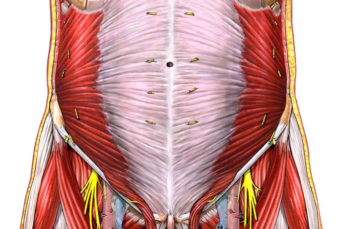
    The internal oblique is just below, or beneath the surface, of the external oblique. The fibers of the internal oblique run perpendicular to the external oblique; this cross-stitching of the muscles give the internal obliqueoblique a tremendous amount of stability and are extremely useful to the respiratory system in breathing and providing support for the organs while they move with the expansion of the lungs. You can see the difference between the internal and external obliques on the photo to the right; the external oblique is located superficially, or closer to the skin, than the internal oblique.

    The origin points for the internal obliques are the thoracolumbar fascia along the spine, the front of the hip plate, or iliac crest, and the bottom of the hip plate along the inguinal ligament. The insertion points are the linea alba, the concave vertical centerline of the abdomen and the rib cartilage of ribs 8 through 12. It stitches up the front of the midline up and back, away from the ground. This muscle is very active in the retention of Uddiyana Bandha, though probably less active than the transverse abdominus muscle. The muscle covers the belly from the bottom of the ribs to the sitting bones.

    Great ways to activate the internal obliques are with isometric exercises to press the arms against the legs, possibly while lying on the back or standing. The muscle spreads up towards the mid-line, so using the airs to stretch the torso will also help to activate the muscle group. Side plank (Vasisthasana) with your leg lifted, standing back-bends, and locust poses can get these muscles active. Anything where you are reaching up with the arms and a straight spine will activate the torso muscles all the way down to the lower internal obliques. Warrior 3 is a very active pose and is awesome for warming up the sides of the body, as are side angle and half-moon pose. When the body is working in concert, it is most powerful. The breath is the composer.

    The internal oblique is a major muscle for moving internal organs on exhalations and making room for the chest cavity to expand. It, combined with the transverse abdominus muscle and diaphragm, are major muscles you feel while you exhale. The muscle has a second major function, which is to move with the external oblique to creation torsion in the spine. when you lift one shoulder and lower the other, you active the internal and external obliques simultaneously. The muscles work in opposition to keep you and your spine upright so that you can stand and more importantly, walk and run.


    The Anatomy of the Abdomen (part 2/4: Internal Obliques) Read More »

    external oblique

    Anatomy of the Abdomen (part 1/4: External Oblique)

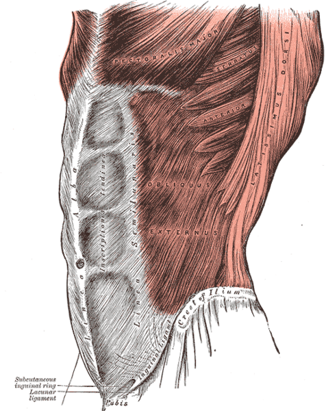
    The abdominal wall consists of four distinct muscles, the transverse abdominus, the internal obliques, the rectus abdominus, and the external obliques. These muscles form three distinct layers, with the rectus abdominus and external obliques on top. The transverse adbominus forms a kind of belt around the lower spine and the internal obliques stitch together up towards the ribs while the external obliques stitch together down from the serratus muscles to the top of the iliac crest.

    The external obliques interlace with the intercostal muscles to support movement of the ribs and spine. They are most important for rotation of the thoracic spine and is what allows for stabilization while twisting and for support during flexion.

    The insertion of the upper fibers occurs at the 5th through 9th ribs where they interlace with the serratus muscles and the 9th through 12th ribs interlacing with the latimus dorsi. The origin of the muscle is at the linea alba, the centerline of the abdomen and the front portion of the iliac crest and the upper part of the pubic tubercle. The external oblique is the largest of the three muscles on the outside of the abdomen.

    The muscle fibers of the external oblique run inwards, towards one another, and down towards the iliac crest at the outside of the hips. origin of the external obliqueThe muscle has eight muscular peaks for each of the eight ribs that it crosses over. The oblique runs down to the Iliac Crest where it inserts into the top of the hip plate and forms the inguinal ligament, which connects the top of the hip crest to the pubis bone at the bottom of the pelvis.

    You activate the external oblique in side plank and stretch them in chaturangas, but there are enormous amounts of exercises to target the muscle. The next layer of abdominal muscles just underneath the external oblique is the internal oblique, which will be part 2. Check back soon to see part 2!

    Anatomy of the Abdomen (part 1/4: External Oblique) Read More »

    foot anatomy

    Foot Anatomy and Function | पाद | pāda

    The Human’s Foot Anatomy

    Consciously, the foot might be the most forgotten part of the human body. At least in my life, my feet are pretty abused, especially when I spent nine months in Paris or 6 months in India. But it’s okay; your foot’s anatomy is designed to take a beating. Being one of the few bi-ped mammals, human feet are simultaneously flexible, sturdy, and versatile. The foot anatomy provides us with the ability to run, jump, climb, grasp, and even use them as a weapon. Our feet are pretty important; they allow us to explore, climb mountains, jump from cliffs, swim, sprint, jump, climb trees, and so much more. Let’s take a look at the anatomy behind one of the most important structural foundations of the body, the human foot.

    Bones of the Foot

    top view of bones of the foot

    Between the ankle and foot anatomies, there are over a hundred tendons, ligaments, and muscles and has over 33 joints. There are 26 bones in the foot and ankle, including 4 metatarsals, the metatarsus, and 5 of the proximal phalanges, middle phalanges, distal phalanges, 3 cuniform bones that form the base of the ankle, the cuboid bone, the navicular, the calcaneus, and the talus. The bones are all very intricate and articulate together to allow the ankle to flex and extend with the foot. The foot’s anatomy, you will find, is intricately related to the ankle, toes, and legs. The arch of the foot is formed from the navicular bone, 3 cuniform bones and the cuboid bone which create a shock absorber in the arch. The talus is the bone that forms the majority of the ankle and the calcaneus is the bone of the heel, while the metatarsals and phalanges form the toes and front of the foot.

    Ligaments of the Foot

    ligaments of the ankle
    medial_foot_ligaments

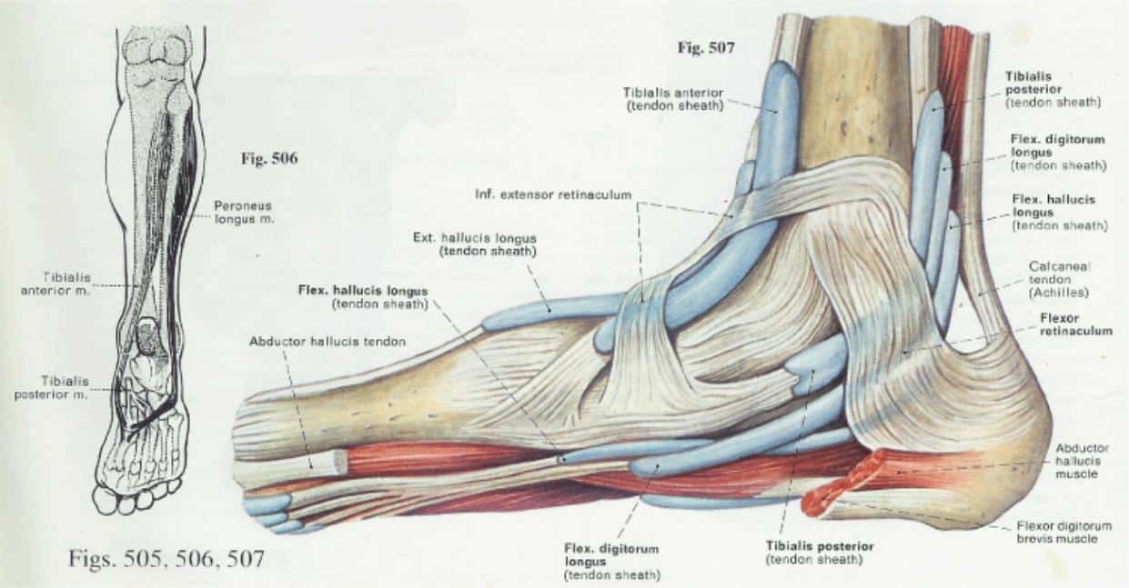
    The ligaments of the foot anatomy are even more complex. The foot is really an intricate array of ligaments and muscles that have multiple cross sections for stability. The ankle has a good amount of study ligaments, with the toes having less and having more muscle and fascia to create flexible support. The major ligaments are the extensor hallicus brevis and longus, the extensor digitorum brevis and longus, the flexor hallicus longus, and the flexor digitorum longus, most of which have tendon sheaths on the outside of the foot.  The peroneus tendons extend up the ankle from the outside of the foot anatomy, the peroneal retinaculum ligaments wrap the back of the foot and the extensor retinaculum ligaments wrap the front of the ankle.

    The Major Muscles of the Foot

    foot_muscles
    veins of the foot

    The major muscles of foot anatomy are the lumbrical muscles, the quadratus plantae muscles, the adductor hallucis muscle, the fibularis longus muscle, and the plantar interosseous muscles. They run along the ligaments to provide extension and flexion for the toes and provide padding for the foot. Fascia covers the muscles and ligaments to provide even more support and a flexible absorption system for the foot to support the rest of the body. Veins flow in between the metatarsals to the insides of the toes. They also criss-cross on the bones of the ankle and run up the ankle to the thighs via the posterior and anterior tibial veins.

    The Nerves of the Foot

    nerves_of_ foot

    The final piece of the foot anatomy is the nervous flow through the ankle and into the toes. The deep fibral nerve runs down the ankle and splits into the peroneal nerves, the medial plantar nerve, the plantar digital nerves, the sural nerve, and the cutaneous nerves. There is a split into 9 nerve endings on the outside and inside of each toe. These nerves are not very sensitive, but are easily mis-aligned to cause significant pain.

    It is kind of easy to see how the feet are often forgotten; their intricacy isn’t very apparent, but the sturdy foundation for human movement is nonetheless very important in our lives. So give your feet a break and show them some love, stretch them out in yoga, happy toes are important to having a well-functioning foot.

    Anatomical Efficiency of the Foot

    Using the ball of the foot is the most essential part of walking, running, and physical exertion. Many people get into the habit of walking on their heels, which can have very negative physiological effects, especially over long periods of time.

    I bought Vibram’s a while back and I love them, but they were sued for false advertising because they really don’t have a positive effect on the foot. I still enjoy being able to grip with my toes, but keep in mind that your foot has natural padding in it; you don’t necessarily want to be wearing extreme support all the time. Try some minimalism, it feels great. Get a foot massage next chance you get, you’ll find that there aren’t many places in your body that can have more stress and usage than your feet.

    Foot Anatomy is complex, what questions do you have? I would welcome any stories about issues with your foot or ankle and how severely it impacted your life.

    Additional Resources:

    Foot Anatomy Animation

    Wikipedia

    Great Pictures of Foot Anatomy

    Foot Anatomy and Function | पाद | pāda Read More »

    Shoulder Anatomy and Physiology

    Human Shoulder Anatomy

    Understanding shoulder anatomy can help to avoid injury, promote rehabilitation, and can assist you in using the joint optimally. Please let me know if you have questions about this article in the comments section at the bottom of the page.

    The human shoulder is a powerful and large anatomical structure. The hinging ball and socket joint allows for vast gains in momentum over short periods of time and is relatively versatile. The shoulder anatomy allows for many types of throwing, fine motor movement down to typing, powerful grasping, hefting objects, climbing, combat, quadruped movement, etc. The shoulder also has a large range of motion; however, this makes the shoulder prone to injury.

    Bones of the Shoulder

    shoulder anatomy bones

    The shoulder joint is relatively loose. There are three main bones of the shoulder: the collarbone, shoulder-blade, and the upper arm bone. These are known as the clavicles, scapula, and humerus, respectively. The shoulder blade also has a bone called the caracoid process which connects to the biceps at the front of the arm and an upwardly angled bone called the acromion that connects to the Clavicle (collarbone) via the CA ligament.

    Ligaments of the Shoulder

    There are large amounts of ligaments and tendons in the shoulder joint, because of its versatility, stability, and strength. As you can see, the three bones of the joint are combined together with vast arrays and webbings of ligaments that allow for the large range of motion while keep the joint stable. Honestly, the joint is so complex that using words to describe it become somewhat useless. So here’s a huge blown up picture for you to look at in awe of how fucking amazing your shoulders are:

    Shoulder_Anatomy_detailed

    Muscles of the Shoulder

    shoulder anatomy muscles

    All of the deep ligaments that you see above are supported by muscles tissue. The muscles that make up the rotator cuff are the infraspinatus, supraspinatus, subscapularis, and teres. There are also three deltoid muscles on the head of the humerus, the rhomboids that connect the shoulder to the spine and the traps which connect the shoulder and neck, and provide support for the shoulder blades.

    The infraspinatus muscle runs along the scapula (shoulder blade), covering the back of it over the teres minor muscle. The teres minor connects the outer arm with the outer lower edge of the shoulder blade. The supraspinatus connects the head of the humerus (arm bone) to the inside edge of the scapula articulating underneath the clavicle (collarbone). The teres major connects the outer clavicle with the back of the humerus; it is more superficial and larger than the teres minor. The subscapularis muscles run on the inside of the shoulder blade, but is not connected to the rib cage which is part of what allows the shoulder blade to have such a broad range of motion. Over all of these muscles are the deltoids, which are the most superficial shoulder muscles. They are separated into anterior, lateral, and posterior sections.

    Nerves of the Shoulder

    The nerves of the shoulder are also complex; consider that the fine motor function of typing must travel from your spine to your fingertips through the intricacies of the shoulder joint. You also have a very responsive feedback loop between your eyes and hands, which travels within the shoulder and into the forearms and fingers.

    There are three primary nerves in the arm that run through the interior of the joint and connect to the digits (fingers).

    shoulder anatomy nerves

    The radial nerve provides innervation to the dorsal muscles of the arm: triceps, extrinsic extensors of the hands, as well as sensory innervation to the back of the hand, except for the pinky and half of the ring finger. It originates from the brachial plexus, carrying fibers from the ventral roots of spinal nerves C5, C6, C7, C8 & T1.

    shoulder_anatomy_brachial

    The ulnar nerve provides innervation to the back of the other three fingers, including the thumb. It also provides the majority of the innervation of the forearm and head of the bicep. The ulnar nerve originates from the C8T1 nerve roots (and occasionally carries C7 fibers) which form part of the medial cord of the brachial plexus, and descends on the posteromedial aspect of the humerus.

    The medial nerve provides innervation for the inside of the thumb, pointer, middle, and half of the ring finger. It also innervates the lateral and inferior portions of the forearm. The median nerve originates from the lateral and medial cords of the brachial plexus, and has contributions from ventral roots of C5-C7(lateral cord) and C8 & T1 (medial cord).

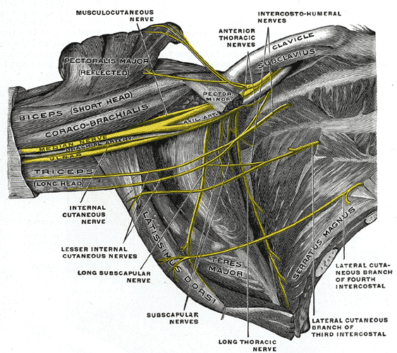
    Here is a final picture of the brachial plexus to assist in visualizing how the nerves flow down the arms.

    This concludes my article on shoulder anatomy; please write any questions below!

    Shoulder Anatomy and Physiology Read More »

    Scroll to Top

    Subscribe

    Get email updates of new articles when they are published

    We promise we’ll never spam! Take a look at our Privacy Policy for more info.